Nikon Corporation Healthcare Business Unit | Asia, Oceania & Middle East
- en Change Region
- Global Site
Image courtesy of Laurence Pelletier Lab, LTRI
June 2021
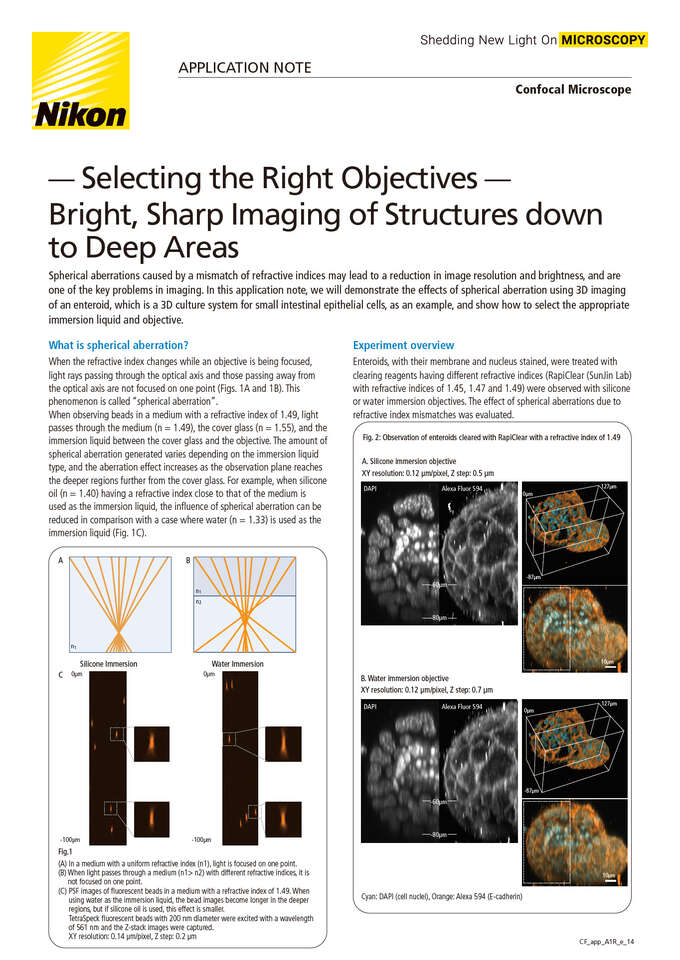
Spherical aberrations caused by a mismatch of refractive indices may lead to a reduction in image resolution and brightness, and are one of the key problems in imaging. In this application note, we will demonstrate the effects of spherical aberration using 3D imaging of an enteroid, which is a 3D culture system for small intestinal epithelial cells, as an example, and show how to select the appropriate immersion liquid and objective.
June 2021
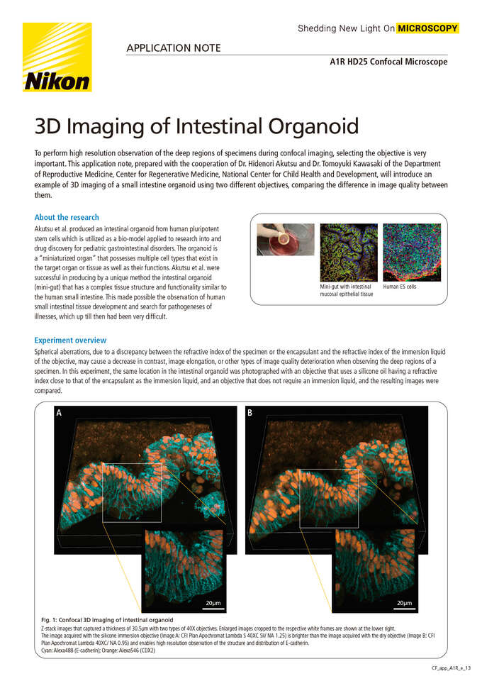
To perform high resolution observation of the deep regions of specimens during confocal imaging, selecting the objective is very important. This application note, prepared with the cooperation of Dr. Hidenori Akutsu and Dr. Tomoyuki Kawasaki of the Department of Reproductive Medicine, Center for Regenerative Medicine, National Center for Child Health and Development, will introduce an example of 3D imaging of a small intestine organoid using two different objectives, comparing the difference in image quality between them.

January 2021
Organs-on-chips more faithfully recapitulate the 3D architectural and functional complexity of native tissues compared to standard 2D tissue culture systems. Yet these advanced cell culture platforms present technical challenges for imaging-based applications. This Application Note demonstrates how the Nikon A1R HD25 confocal point-scanning system, CFI S Plan Fluor LWD 20XC objective and NIS-Elements software can enable rapid, deep, quantitative imaging of living cells in the Emulate Organ-Chip platform.
February 2019
Capturing the dynamics of living systems requires high acquisition rates. Large samples, such as whole model organisms, additionally require a large field of view. The Nikon A1R HD25 confocal system provides both, combining Nikon’s improved HD high speed resonant scanner with an unprecedented 25 mm field of view. The performance of this system is evaluated in zebrafish embryos.
November 2018
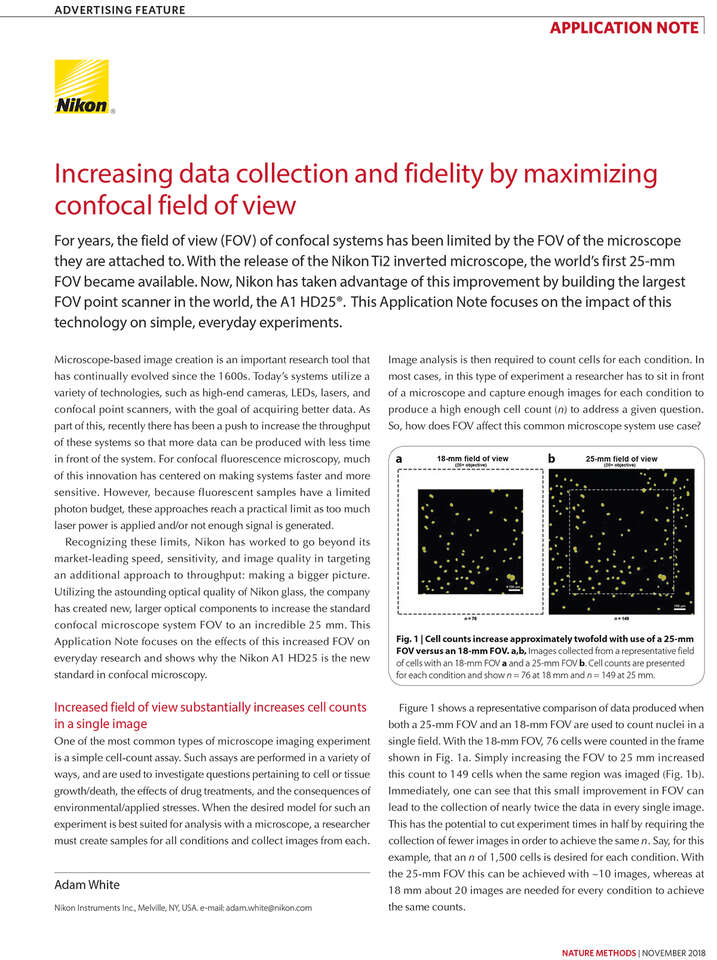
For years, the field of view (FOV) of confocal systems has been limited by the FOV of the microscope they are attached to. With the release of the Nikon Ti2 inverted microscope, the world’s first 25-mm FOV became available. Now, Nikon has taken advantage of this improvement by building the largest FOV point scanner in the world, the A1 HD25. This Application Note focuses on the impact of this technology on simple, everyday experiments.


