Nikon Instruments Inc. | Americas
- en Change Region
- Global Site
August 2023
In the field of brain research, multiphoton confocal microscopy is necessary for understanding the mysterious functions and complex circuitry of the brain. The brain is the nexus that controls our thoughts, emotions and actions, and clarifying the exact mechanisms thereof may lead to the treatment of neurological disorders and improvement of cognitive functions. This application note introduces an example of super-resolution imaging for capturing the myelin in a mouse brain using the AX R MP with NSPARC multiphoton confocal microscope, equipped with a super-resolution detector.
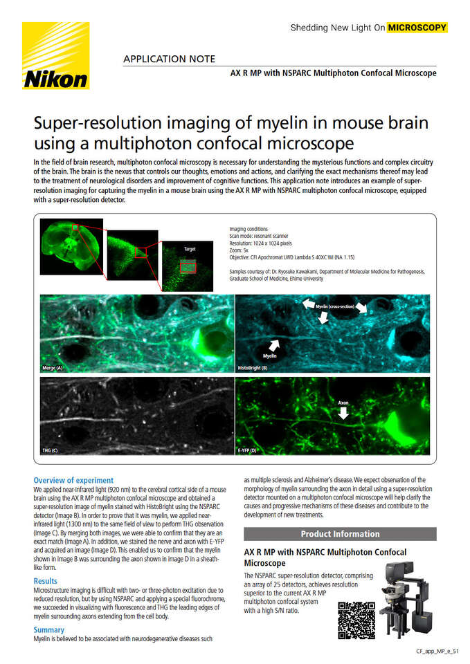
We applied near-infrared light (920 nm) to the cerebral cortical side of a mouse brain using the AX R MP multiphoton confocal microscope and obtained a super-resolution image of myelin stained with HistoBright using the NSPARC detector (Image B). In order to prove that it was myelin, we applied near-infrared light (1300 nm) to the same field of view to perform THG observation (Image C). By merging both images, we were able to confirm that they are an exact match (Image A). In addition, we stained the nerve and axon with E-YFP and acquired an image (Image D). This enabled us to confirm that the myelin shown in image B was surrounding the axon shown in image D in a sheath-like form.
Imaging conditions
Scan mode: resonant scanner
Resolution: 1024 x 1024 pixels
Zoom: 5x
Objective: CFI Apochromat LWD Lambda S 40XC WI (NA 1.15)
Samples courtesy of: Dr. Ryosuke Kawakami, Department of Molecular Medicine for Pathogenesis, Graduate School of Medicine, Ehime University
Merge (A)
HistoBright (B)
THG (C)
E-YFP (D)
Microstructure imaging is difficult with two- or three-photon excitation due to reduced resolution, but by using NSPARC and applying a special fluorochrome, we succeeded in visualizing with fluorescence and THG the leading edges of myelin surrounding axons extending from the cell body.
Myelin is believed to be associated with neurodegenerative diseases such as multiple sclerosis and Alzheimer's disease. We expect observation of the morphology of myelin surrounding the axon in detail using a super-resolution detector mounted on a multiphoton confocal microscope will help clarify the causes and progressive mechanisms of these diseases and contribute to the development of new treatments.


