Nikon Europe B.V. | Europe & Africa
- en Change Region
- Global Site
March 2025
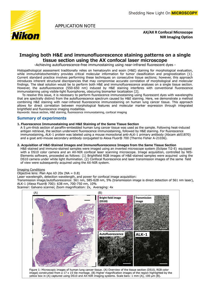
Histopathological assessment traditionally relies on hematoxylin and eosin (H&E) staining for morphological evaluation, while immunohistochemistry provides critical molecular information for tumor classification and prognostication [1]. Current standard practice involves performing these techniques on consecutive tissue sections; however, this approach introduces inherent structural discrepancies that may compromise accurate correlation of morphological and molecular findings. The ideal solution would be to perform both H&E and immunofluorescence analyses on a single tissue section. However, the autofluorescence (550-650 nm) induced by H&E staining interferes with conventional fluorescence immunostaining using visible-light fluorophores, obscuring biomarker localization [2].
To resolve this issue, it is necessary to perform fluorescence immunostaining using fluorescent dyes with wavelengths that are spectrally distinct from the autofluorescence spectrum caused by H&E staining. Here, we demonstrate a method combining H&E staining with near-infrared fluorescence immunostaining on human lung cancer tissue. This approach allows for direct correlation between morphological features and molecular marker expression through integrated brightfield and fluorescence imaging modalities.
Keywords: tissue section, H&E staining, fluorescence immunostaining, confocal imaging