Nikon
Nikon Healthcare: A Century of Microscopy
Nikon Europe B.V. | Europe & Africa
  • en Change Region
  • Global Site
  • Artboard 1

Select Your Region and Language

Products and Promotions may differ based on your selected Region.

Americas

  • en English
  • es Español
  • fr Français
  • pr Português

Europe & Africa

  • en English
  • de Deutsch
  • es Español
  • fr Français
  • it Italiano
  • pr Português

Japan

  • ja 日本語

Korea

  • ko 한글

Chinese Mainland

  • zh 简体中文

Asia, Oceania & Middle East

  • en English

Return to the top of the page

  • Products
  • CRO
  • Applications
  • Resources
  • BioImaging Centers
  • About Us
  • Contact

Microscope Products

Light Microscopes

  • Inverted Microscopes
  • Upright Microscopes
  • Polarizing Microscopes
  • Stereo Microscopes

Advanced Imaging Systems

  • Confocal and Multiphoton Microscopes
  • Super-Resolution Microscopes
  • Slide Scanning
  • Photostimulation & TIRF

Digital Microscopes

Software

Cameras

Optics

Accessories

OEM

Discontinued Products

Nikon BioImaging Labs

Nikon BioImaging Labs provide contract research services for microscope-based imaging and analysis to the biotech, pharma, and larger research communities. Each lab's full-service capabilities include access to cutting-edge microscopy instrumentation and software, but also the services of expert biologists and microscopists, who are available to provide quality cell culture, sample preparation, data acquisition, and data analysis services.

Boston, United States

Europe

Japan

Applications

Life Sciences

  • Deep Learning in Microscopy
  • Drug Discovery
  • Electrophysiology
  • Live Cell Imaging
  • Microinjection
  • Nano-Scale Imaging
  • Organoid and Organ-on-a-chip Imaging
  • Regenerative Medicine
  • Tissue Culture
  • Whole Organism and Tissue Imaging

Clinical Research

  • Assisted Reproductive Technology (ART)
  • Gout Testing
  • Imaging Fixed Slides

Education

OEM

Resources

Featured Resources

  • Latest News & Updates

    Sign up to our mailing list to be first to hear about our new products and exclusive offers!

  • PubScope

    Search and filter over 125,000 Open Access Articles that utilize Nikon products and supported third party systems.

  • Resolution Calculator

    Input appropriate parameters to calculate important performance specifications such as resolution, depth of field, sampling rate, confocality, and more.

  • Software/Firmware Downloads

    Download software and firmware programs for Nikon microscope products.

  • Online Guides

    Step-by-step guides for aligning and operating select Nikon microscopes.

  • MicroscopyU

    Nikon's MicroscopyU is a top source for educational information about optical microscopy.

  • Webinars

    Webinars related to optical microscopy technology and its applications.

  • Objective Selector

    Find the right Nikon objective to fit your workflow.

Browse Resources

Support

About Us

  • News
  • Company Profile
  • Careers
  • Service
  • Sustainability
  • Modern Slavery Act Statement
  • Home
  • CRO
  • Boston, USA
  • Applications
  • mRNA Quantification (smFISH) for Gene Expression Analysis

Nikon BioImaging Labratories

Boston, USA

  • Europe
  • Japan
  • Overview
  • Applications
  • Research Services
  • Expertise
  • FAQs
  • Resources
  • News & Events

mRNA Quantification (smFISH) for Gene Expression Analysis

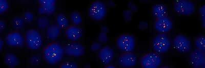
Single molecule fluorescence in situ hybridization (smFISH), known for its ability to quantify individual RNA molecules, is widely used to characterize spatio-temporal patterns of gene expression in single cells. At NBIL we harness this technique to provide RNA quantification services for research applications such as gene expression analysis in response to drug candidates, spatial biology studies, and more.

Overview of smFISH
Raw Image
Processed and segmented image
Data analysis output

Challenges

Fundamentally, smFISH relies on quantification of small, bright spots within the cell. The quality of data and subsequent conclusions depends on the quality of the acquired images, which is hindered by low fluorescence signals, high fluorescence backgrounds, and proximity of adjacent spots. Furthermore, it is important to segment the spots for analysis in a consistent manner and manage potentially large quantities of data depending on experiment complexity.

Solutions

Nikon’s diverse lineup of world-renowned objective lenses are designed to optimally image a huge variety of sample types. Acquiring clear images enables powerful, repeatable image analysis. The NIS-Elements software is equipped with a full toolbox of image processing, segmentation, and analysis options, including AI-driven functions for pre-defined and customizable feature identification. It is also possible to develop hardware routines to increase imaging throughput and complexity.

The NBIL is staffed with expert assay scientists & microscopists who will work with you to develop and execute customized image acquisition and analysis workflows to advance your research.

  • Home
  • CRO
  • Boston, USA
  • Applications
  • mRNA Quantification (smFISH) for Gene Expression Analysis
Nikon
  • FacebookFacebook
  • LinkedInLinkedIn
  • XX
  • InstagramInstagram

Contact Nikon Europe B.V.

Stroombaan 14, 1181 VX Amstelveen
The Netherlands

Enquiries Tel: 0031-(0)-20-7099-000

About Us

  • News
  • Events
  • Company Profile
  • Careers
  • Service
  • Sustainability
  • Well-being
  • Modern Slavery Act Statement

Popular Links

  • Latest News & Updates
  • Objective Selector
  • Resolution Calculator
  • PubScope
  • OEM
  • Nikon Small World
  • MicroscopyU

Other Nikon Products

  • Imaging Products
  • Industrial Metrology
  • Semiconductor Lithography Systems
  • FPD Lithography Systems
  • Contact
  • Site Map
  • Privacy
  • Cookie Policy
  • Terms of Use
  • Careers

© 2025 Nikon Europe B.V.