Nikon Europe B.V. | Europe & Africa
- de Change Region
- Global Site
Mai 2025
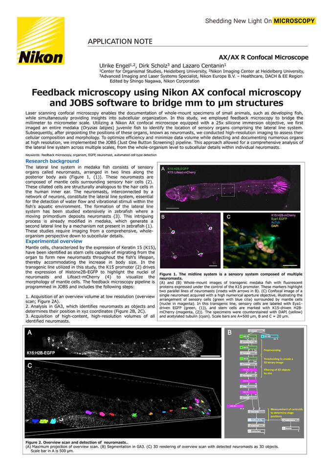
Laser scanning confocal microscopy enables the documentation of whole-mount specimens of small animals, such as developing fish, while simultaneously providing insights into subcellular organization. In this study, we employed feedback microscopy to bridge the millimeter to micrometer scale. Utilizing a Nikon AX confocal microscope equipped with a 25x silicone immersion objective, we first imaged an entire medaka (Oryzias latipes) juvenile fish to identify the location of sensory organs comprising the lateral line system. Subsequently, after pinpointing the positions of these organs, known as neuromasts, we conducted high-resolution imaging to assess their cellular composition and morphology. To optimize efficiency and minimize data volume while detecting and documenting numerous organs at high resolution, we implemented the JOBS (Just One Button Screening) pipeline. This approach allowed for a comprehensive analysis of the lateral line system across multiple scales, from the whole-organism level to subcellular details within individual neuromasts.
Keywords: feedback microscopy, organism, EGFP, neuromast, automated cell type detection