Nikon Instruments Inc. | Americas
- en Change Region
- Global Site
November 2023
Motor nerves are a collection of many axons assembled and aligned in one direction from the spinal cord to muscles. The fasciculation of axons provides them guidance, strength, and a healthy microenvironment. Therefore, it is of great significance to develop an in vitro model that mimics in vivo motor neuron properties and reproduces its structure.
Professor Yoshiho Ikeuchi et al., Institute of Industrial Science, The University of Tokyo, aims to reproduce the physiological environment of the human body using a microfluidic device.
This application note introduces an example of the spontaneous formation of motor nerve organoids in a microchip embedded in a 96-well plate, captured using a confocal microscope
Keywords: microdevices, motor nerve organoids, long-term time-lapse imaging, confocal.
Figure 1. Motor nerve organoid formation in a microdevice
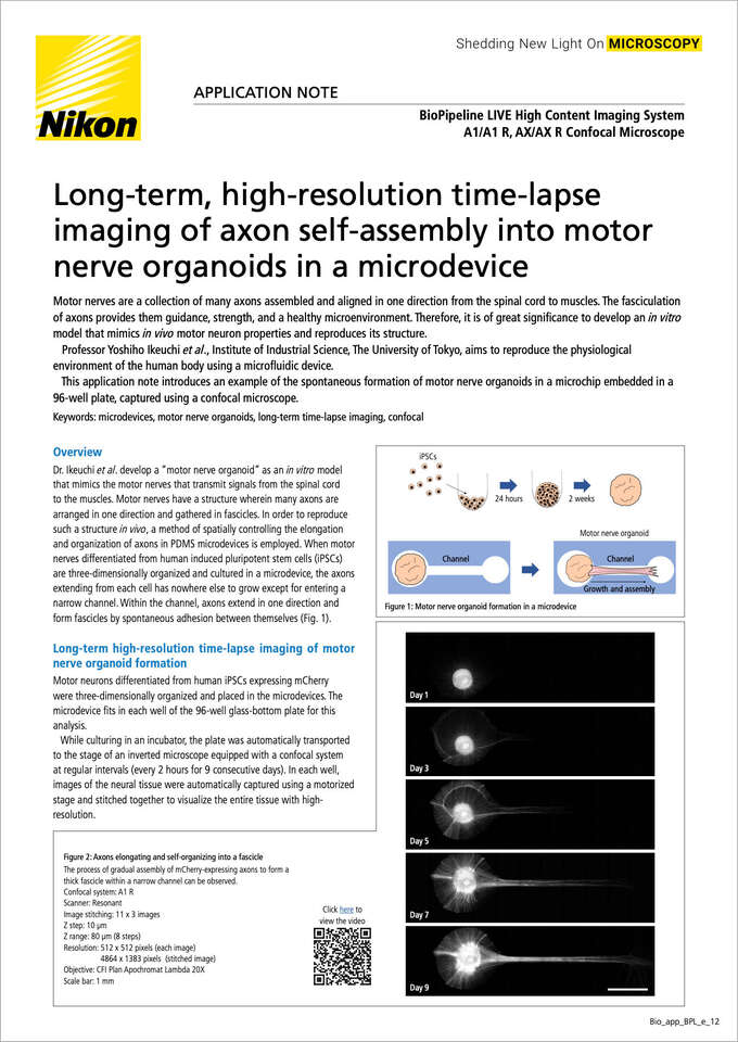
Dr. Ikeuchi et al. develop a "motor nerve organoid" as an in vitro model that mimics the motor nerves that transmit signals from the spinal cord to the muscles. Motor nerves have a structure wherein many axons are arranged in one direction and gathered in fascicles. In order to reproduce such a structure in vivo, a method of spatially controlling the elongation and organization of axons in PDMS microdevices is employed. When motor nerves differentiated from human induced pluripotent stem cells (iPSCs) are three-dimensionally organized and cultured in a microdevice, the axons extending from each cell has nowhere else to grow except for entering anarrow channel. Within the channel, axons extend in one direction and form fascicles by spontaneous adhesion between themselves (Fig. 1).
Motor neurons differentiated from human iPSCs expressing mCherrywere three-dimensionally organized and placed in the microdevices. The microdevice fits in each well of the 96-well glass-bottom plate for this analysis.
While culturing in an incubator, the plate was automatically transported to the stage of an inverted microscope equipped with a confocal system at regular intervals (every 2 hours for 9 consecutive days). In each well, images of the neural tissue were automatically captured using a motorized stage and stitched together to visualize the entire tissue with high-resolution.
Figure 2. Axons elongating and self-organizing into a fascicle
The process of gradual assembly of mCherry-expressing axons to form a thick fascicle within a narrow channel can be observed.
Confocal system: A1 R
Scanner: Resonant
Image stitching: 11 x 3 images
Z step: 10 μm
Z range: 80 μm (8 steps)
Resolution: 512 x 512 pixels (each image)
4864 x 1383 pixels (stitchedimage)
Objective: CFI Plan Apochromat Lambda 20X
Scale bar: 1 mm
Figure 3. Self-organization process of axons within a channel and a spheroid chamber
(a) inside channel, (b) near spheroid containing cell bodies
Scale bar: 200 μm
Objective: CFI Plan Apochromat Lambda 20X
Figure 4. Motor nerve organoid with axons self-organized into a fascicle in a microdevice in a well
(Left) Microdevice placed in a well of a 96-well plate
(Center) Motor nerve organoid with extended axons in the microdevice
(Right) Motor nerve organoid extracted from the microdevice
It takes more than a week for axonal elongation and self-organization of motor nerves derived from human iPSCs. However, the entire process could be observed seamlessly using the BioPipeline LIVE microscope automated incubation and imaging system, which combines an incubatorwith a confocal microscope (Figs. 2 and 3). High-resolution, long-term time-lapses revealed the process by which countless axons undergo complex interactions and ultimately self-organize into fascicles (Figs. 2 and 3).
BioPipeline LIVE, a microscope automated incubation and imaging system that combines incubation and confocal microscopy, enables both long-term time-lapse imaging and high-resolution imaging.
The observations in this study revealed that radially extending axons and gathering axons in an arc are entwined near the cell body, and that all the axons eventually extend in the direction of the channel (Fig. 3b). In addition, when the axonal fascicle is formed in the channel, bright spots on the axon gather while being attracted toward the cell body (Fig. 3a). The physiological significance of this phenomenon and its mechanism require further detailed investigation.
In order to observe fine axons in thick, multilayered tissue, it is necessary to observe a wide area with high resolution in three dimensions. The confocal microscope within BioPipeline LIVE can easily acquire stitched images and perform Z-stack imaging. Since this system can automatically analyze many 96-well plates, it is also applicable to compound and drug screening.
We would like to express our sincere gratitude to Dr. Yoshiho Ikeuchi and Tomoya Duenki of Institute of Industrial Science, The University of Tokyo, for their generous cooperation in preparing, providing, and imaging specimens.
Using Ti2-E inverted research microscope as a platform, this system offers a wide range of objectives, imaging devices and observation methods, and can be also combined with a confocal system. The incubator can hold up to 44 well plates.


