
Smart Automation with Confocal Microscope: ECLIPSE Ji Changes the Future of 3D Cell Research
AdHuCell platform and Research Institute for Biosciences
University of Mons - UMONS
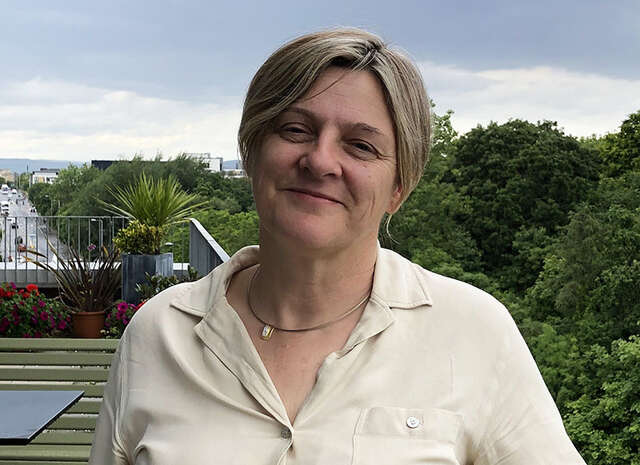
Professor Sylvain Gabriele, SYMBIOSE Lab
Professor Sylvain Gabriele at the University of Mons (UMONS), Belgium, is a leading researcher in mechanobiology, investigating how cells sense, respond to, and retain memory of mechanical forces.
In this interview, we spoke with Professor Gabriel about how the Nikon ECLIPSE Ji-AX Confocal Integrated System has advanced their understanding of complex 3D biological systems.

시간을 초월한 성능 - LIV의 혁신을 뒷받침하는 장기 사용 현미경
크리스티안 콘체 박사
함부르크 라이프니츠 바이러스학 연구소 (LIV) 광학현미경·이미지 분석 기술 플랫폼 부문장
니콘 현미경 사업 100주년을 기념하여, 'Enduring Excellence(시간을 초월한 성능)' 시리즈에서는 니콘 시스템을 오랜 세월에 걸쳐 활용하며, 장비에 담긴 지속가능성과 장기적 가치를 몸소 실증하고 있는 연구자들을 소개합니다. 이번 편에서는 함부르크 라이프니츠 바이러스학 연구소 (LIV) Nikon Center of Excellence 에서 광학현미경·이미지 분석 기술 플랫폼 부문장을 맡고 있는 크리스티안 콘체 씨를 만났습니다. 콘체 씨는 연구자로서의 커리어 전반에 걸쳐 장기간 사용되어 온 니콘 현미경을 활용해 왔습니다.

“이 현미경은 배아연구원이 하루에 더 많은 ICSI/IMSI 시술을 수행할 수 있도록 돕고 배아의 질 향상에도 기여할 것입니다”
데이비드 하레뇨 마르티네즈 박사
CHIREC IVF 연구소의 선임 배아생물학자
벨기에 생식의학회(BSRM) 이사회 임원
유럽에서도 의료 선진국으로 손꼽히는 벨기에의 CHIREC(Centre Hospitalier Interrégional Edith Cavell) 병원 그룹은 다양한 전문 분야에서 포괄적이고 혁신적인 의료 서비스를 발전시키기 위해 노력하고 있습니다.
벨기에는 세계 최초로 ICSI(세포질 내 정자 주입) 시술을 성공적으로 수행해 낸 나라로도 알려져 있습니다*. CHIREC의 IVF(체외 수정) 연구소는 이러한 혁신 정신을 바탕으로 환자 개개인에게 맞춘 정교한 난임 치료 서비스를 제공합니다.
또한 ICSI/IMSI 시술용으로 설계된 니콘의 전동 도립 현미경 ECLIPSE Ti2-I는 이러한 임상 환경에 새로운 가능성을 열어줄 것으로 기대됩니다.
본 인터뷰에서는 CHIREC IVF 연구소에서 16년 넘게 근무해온 선임 배아생물학자 데이비드 하레뇨 마르티네즈 박사님을 만나 현미경의 광학 기능과 자동화 기능에 대한 소감과 함께, 이러한 기술이 치료 결과에 미칠 잠재적 영향력에 관해 이야기를 나누었습니다.
*세계 최초의 ICSI 시술은 UZ 브뤼셀에서 성공적으로 이루어졌습니다.
https://brusselsmorning.com/thirty-years-ago-the-very-first-icsi-baby-was-born-in-uz-brussel/20731/
면책 고지: 본 기사에는 니콘 제품에 대한 의료 전문가의 의견이 포함되어 있습니다. 그러나 이것이 제품의 효능, 효과 또는 성능을 보장하지 않으며 의료 전문가가 해당 제품을 보증, 추천 또는 선택함을 의미하지도 않습니다.

시간을 초월하는 성능 - VIB-KU Leuven에서의 실제 사례
니키 코르타우트(Nikky Corthout)
광학 현미경 전문가 - VIB 바이오이미징 코어 루벤, VIB-KU 루벤 뇌질환센터
파블로 에르난데스 바라스(Pablo Hernández Varas)
코어시설 책임자 - VIB 바이오이미징 코어 루벤, VIB-KU 루벤 뇌질환센터
100년 이상 동안 니콘의 현미경 시스템은 장기 사용을 전제로 설계되어 왔습니다. 이번에는 벨기에 루벤 가톨릭대학교(KU Leuven)에 설치된 Nikon Center of Excellence 내 VIB 바이오이미징 코어 루벤에서 활동하는 파블로 에르난데스 바라스와 니키 코르타우트를 만나 이야기를 들었습니다. 두 분은 17년 이상 동일 연구실에서 사용되어 온 니콘 A1 R 공초점 현미경 시스템을 활용하고 있습니다. 그들의 경험을 통해 시스템의 내구성뿐만 아니라 현재도 최첨단 과학적 발견을 지원하고 있는 모습을 소개합니다.

“자동화된 설정으로 장시간에 걸친 실습에도 손목과 팔의 부담이 줄어 더욱 쾌적해졌습니다”
한국의 보조 생식 기술 아카데미에서 배아 연구원 양성에 기여하는 ECLIPSE Ti2-I
김은경 박사
CHA UNIVERSITY MEDICAL CENTER
Global CHA Embryolab Academy
난임 트레이닝 센터실 난임 TQM실장
전 세계적으로 주요한 과제 중 하나인 저출산 문제를 해결하기 위해서는 난자와 정자, 수정란(배아)을 다루는 배아 연구원의 양성이 무엇보다 시급합니다. 이에 한국의 CHA 병원・바이오 그룹은 보조 생식 기술을 뒷받침하는 배아 연구원의 체계적인 양성을 위해 Global CHA Embryolab Academy(GCEA)라는 본격적인 교육 기관을 설립했습니다. 이곳에서 니콘의 ICSI/IMSI용 전동 도립 현미경 ECLIPSE Ti2-I가 활용되고 있습니다. 본 인터뷰에서는 오랜 경험을 지닌 배아 연구원이자, 동 시설의 난임 트레이닝 센터에서 토털 퀄리티 매니지먼트 실장을 맡고 계시는 김은경 박사님께 니콘 현미경의 조작감과 도입 효과 등에 대한 의견을 여쭈었습니다.
※이 페이지에는 니콘 제품에 대한 의료 종사자 인터뷰가 포함되어 있습니다.
이 인터뷰는 제품의 효능, 효과 또는 성능을 보장하지 않으며 의사가 해당 제품을 보증, 추천 또는 선택한다는 것을 의미하지 않습니다.

SMZ18을 활용한 샘플 작성이 이미지 촬영의 핵심
요네 프로덕션
모리오카 가나코 님 (연구부 이사)
고품질의 샘플을 만드는 것은 아름다운 이미지를 만드는 첫 번째 단계입니다. 현미경 검사를 위한 샘플을 만드는 데는 오랜 시간이 걸리는데 인내심과 기술 그리고 독창성이 필요한 정교한 과정입니다. 생명과학 영상을 제작하는 요네 프로덕션에서는 이처럼 섬세한 샘플을 만들기 위해 실체 현미경 SMZ18을 활용하고 있습니다. 이번 기사에서는 요네 프로덕션에서 샘플 준비를 담당하는 연구부의 모리오카 가나코 님께서 SMZ18을 사용한 경험을 담았습니다.

‘자동화된 설정이 ART 임상의 효율성과 연구 및 개발을 뒷받침합니다’
보조 생식 기술(ART)에 공헌하는 ECLIPSE Ti2-I
코바야시 타츠야 님
후지타 의과대학 도쿄 첨단의료연구센터
재생의료센터 배양실장
후지타 의과대학 의료과학부 연구추진팀
규제과학분야 준교수
생식보조의료관리 배아연구원
사회가 직면한 중요 과제 중 하나인 저출산을 해결하기 위해 보조 생식 기술(ART)의 지속적인 발전이 기대되고 있습니다. 후지타 의과대학 도쿄 첨단의료연구센터에서는 아이를 갖고 싶어 하는 사람들이 한 명이라도 더 많이 부모가 될 수 있도록 ART의 임상과 연구 및 개발에 힘쓰고 있습니다. 이러한 노력에 니콘의 ICSI/IMSI용 전동 도립 현미경 ECLIPSE Ti2-I도 힘을 보태고 있습니다. 본 인터뷰에서는 배아연구원이자 같은 대학 의료과학부의 준교수를 역임하고 있는 코바야시 타츠야 님께 니콘 현미경을 도입하게 된 경위와 실제 사용했을 때의 조작감 등을 물었습니다.
※본 콘텐츠에 당사의 제품이 소개되어 있지만 당사 제품은 일본 국내에서 의료기기가 아닙니다. 또한 당사 제품에 대한 의료 종사자의 코멘트가 기재되어 있으나 그것이 당사 제품의 효능과 효과 및 성능을 보증하지 않으며 해당 의료 종사자가 당사 제품을 공인하거나 추천, 지도 또는 사용하고 있음을 나타내지도 않습니다.

고객 인터뷰 - 안과 진료의 미래에 기여하는 ECLIPSE Si
‘컴팩트한 사이즈, 쉬운 조작 그리고 우수한 화질이 ECLIPSE Si를 선택한 이유입니다.’
인체공학적인 설계로 자연스러운 자세에서 관찰과 효율적인 조작을 추구한 니콘의 생물 현미경 ECLIPSE Si. 에히메현의 이시즈치 안과에서는 결막염 등의 안질환 진료에 안지 도말검사를 도입하여 ECLIPSE Si를 검사에 활용하고 있습니다. 이 현미경을 선택한 이유와 실제 현장에서 느끼는 조작감 등을 이시즈치 안과의 이사장인 스즈키 타카시 박사에게 물었습니다.
※이 페이지에는 니콘 제품에 대한 의사의 인터뷰가 포함되어 있습니다. 이 인터뷰는 제품의 효능, 효과 또는 성능을 보장하지 않으며 의사가 해당 제품을 보증, 추천 또는 선택한다는 것을 의미하지 않습니다.

YOUR SWITCH TO THE FUTURE
스위스 바젤대학교 생물의학과 (DBM)
파스칼 로렌츠
마이클 아반토 박사
니콘 우수 센터장
현미경 코어 시설장
연구 개요: 면역학 및 전염병, 신경과학, 암 생물학, 조직 발달 및 재생.

연구원 인터뷰 - Dr. Kevin Richetin
니콘 현미경 홍보대사인 Dr. Kevin Richetin은 알츠하이머병과 퇴행성 신경질환을 위한 도구 개발을 연구하는 신경과학 연구자입니다. 이 인터뷰 영상에서 Dr. Kevin Richetin은 니콘 현미경을 선택한 이유는 NIS-Elements 소프트웨어를 사용하면 분석과 자동화를 원활하게 통합할 수 있기 때문이라고 설명합니다. 그는 이 고유한 기능이 입자를 매우 높은 해상도로 장기간 추적하는 데 매우 유용하며, 이것이 니콘을 차별화하는 기능이라고 강조합니다.

고객 인터뷰 - 디지털 병리학을 앞당기는 혁신적인 디지털 현미경
키타사토 대학 키타사토 연구소 병원
병리진단과 부장
마에다 이치로 박사
환자 개개인의 병리학적 특성에 따라 최적의 의료를 제공할 수 있는 다양한 치료법이 지금도 개발되고 있습니다. 그러므로 치료 방침과 전략 수립에 필수불가결한 병리 진단의 역할이 그 어느 때보다 중요해지고 있습니다. 그렇지만 일본뿐만 아니라 전 세계적으로도 이러한 역할을 담당할 숙련된 병리학자가 부족하다는 것이 문제가 되고 있습니다. 이 문제를 해결하기 위해 디지털 병리학이 주목받고 있습니다. 이번 기사에서는 키타사토 대학 키타사토 연구소 병원 병리진단과의 마에다 이치로 박사에게 디지털 이미지 디스플레이 광학 현미경 'ECLIPSE Ui'를 사용한 소감을 들어보았습니다.
*마에다 박사는 자신의 개인적인 경험을 바탕으로 이 현미경의 특징과 이 제품이 보여주는 임상적 유용성에 대해 의견을 보내주었습니다.

“난자와 정자 뒤에는 환자분이 있다는 것을 항상 염두에 두며, 그것들은 '사람'이라는 생각으로 다룹니다.”
이에다 쇼코 님
미나토미라이 유메클리닉
배양실 실장
시마무라 스미 님
미나토미라이 유메클리닉
배양실 주임
일본에서는 2022년 4월부터 난임 치료에 대한 보험 적용이 시작되어 임신을 원하는 부부의 현미수정 시도에 대한 부담이 덜어졌습니다. 하지만 일반적으로 아직 '난임은 병이 아니다'라는 인식이 남아 있기 때문에 치료에 대해 깊은 고민과 조급함을 느끼는 환자분들이 많습니다. 그런 마음을 헤아려 아기의 탄생을 돕는 일에 보람을 느낀다는 미나토미라이 유메클리닉의 이에다 쇼코 씨와 시마무라 스미 씨에게 'ECLIPSE Ti2-I'를 사용한 소감을 들어보았습니다.
※본 콘텐츠에 당사의 제품이 소개되어 있지만 당사 제품은 일본 국내에서 의료기기가 아닙니다. 또한 당사 제품에 대한 의료 종사자의 코멘트가 기재되어 있으나 그것이 당사 제품의 효능과 효과 및 성능을 보증하지 않으며 해당 의료 종사자가 당사 제품을 공인하거나 추천, 지도 또는 사용하고 있음을 나타내지도 않습니다.

연구원 인터뷰 - YOUR SWITCH TO THE FUTURE
신시내티 아동병원 의료센터
아동병원(UC) 소아응급실 교수
매튜 코프론 박사
신시내티 낭포성섬유증 치료연구센터
알리샤 오스트만
다케베 연구소 소화기내과
의사 이와사와 겐타로
연구 개요: 아동의 건강 증진. 유사장기 개발. 낭포성 성유증 환자 대상 치료약 연구

연구원 인터뷰 - YOUR SWITCH TO THE FUTURE
유럽 분자영상연구소(EIMI)
생체분자영상학과
프리데만 키퍼 박사
연구 개요: 분자 메커니즘이 생물학적으로 어떻게 인간의 세포와 조직을 구성하는지에 대한 연구. 멀티스케일 이미징 분석.


연구원 인터뷰 - YOUR SWITCH TO THE FUTURE
중국 베이징 소재 중국 뇌연구소
연구책임자
후 자오 박사
연구 개요: 새로운 조직 투명화 기술 개발 및 이를 바탕으로 한 신경망 뇌지도 커넥텀의 맵핑 플랫폼 구축

고객 인터뷰 - "병리학자에 대한 고정관념을 바꿀 수 있다고 믿습니다."
도쿄대학 병원
차세대 병리학 정보 네트워킹학과
교수, 프로젝트 리더, 의학 박사
사사키 다케시
의학 분야에서 병리학자는 관찰을 확인하는 데 매우 중요한 역할을 합니다. 연구원들은 장시간 현미경으로 관찰하는 경우가 많기 때문에 신체적으로나 정신적으로 부담이 큽니다. 니콘은 이러한 스트레스를 완화하려는 목표로 병리학자들이 접안렌즈가 아닌 모니터를 통해 표본의 고해상도 이미지를 볼 수 있는 새로운 병리 검사용 현미경인 ECLIPSE Ui를 개발했습니다. 이 현미경을 평가한 도쿄대학 병원의 다케시 사사키 박사는 이 현미경에 대한 소감을 다음과 같이 말합니다.
*사사키 박사는 자신의 개인적인 경험을 바탕으로 이 현미경의 특징과 이 제품이 보여주는 임상적 유용성에 대해 의견을 보내주었습니다.

Nikon BioImaging Lab contributes to “mini-gut” research
Dr. Hidenori Akutsu
Director of the Department of Reproductive Medicine, Center for Regenerative Medicine, National Center for Child Health and Development
Nikon Equipment and Services

연구원 인터뷰 - YOUR SWITCH TO THE FUTURE
오사카 대학 미생물병 연구소
교수 미키 히로아키
부교수 후나토 요스케
조교수 하시즈메 오사무
환경 응답 연구 부문
세포 제어 분야
연구 개요: 세포 내에서 마그네슘 이온을 배출하는 역할을 담당하는 막단백질 분자 사이클린 M의 기능 해석

ECLIPSE Ei로 교실 학습 지원
“학생들에게 사용 방법을 가르치기 쉽고, 학생들도 사용하기 간단하다고 생각합니다.”
현미경을 활용한 관찰은 과학의 기초를 배우는 데 매우 중요한 역할을 합니다. 니콘의 ECLIPSE Ei는 “처음 조작하는 학생들에게도 사용자 친화적인 현미경”이라는 개념을 바탕으로 교육용 현미경으로 개발되었습니다. 교실에 ECLIPSE Ei가 있는 일본 사이타마현의 릿쿄 니이자 중고등학교에 재직 중인 이즈미 리카 과학 선생님을 만나 ECLIPSE Ei 현미경을 선택하게 된 배경과 이유, 사용자 경험 및 학생들의 반응에 대한 얘기를 들어보았습니다.

ECLIPSE Ci-L plus는 보다 편안한 임상 검사에 도움이 됩니다.
“높은 광학 성능과 함께 사려 깊은 디자인이 일상 업무에 도움이 된다는 것을 깨달았습니다.”
임상 검사는 의료에서 중요한 역할을 합니다. 니콘은 일상적으로 현미경을 사용하는 임상의와 실험실 기술자의 신체적, 정신적 부담을 줄여준다는 개념으로 새로운 생물학적 현미경인 ECLIPSE Ci-L plus를 개발했습니다. 이번 인터뷰에서는 일본 지바현 남부의 대표 병원인 가메다 메디컬 센터 해부병리과의 Akira Yoshikawa 박사에게 이 현미경과 실용적인 일상용으로 새로 개발한 현미경용 대물렌즈 ‘CFI Plan Apochromat Lambda D’ 를 사용한 소감에 대해 이야기를 나눴습니다.
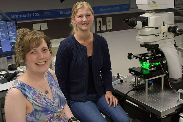
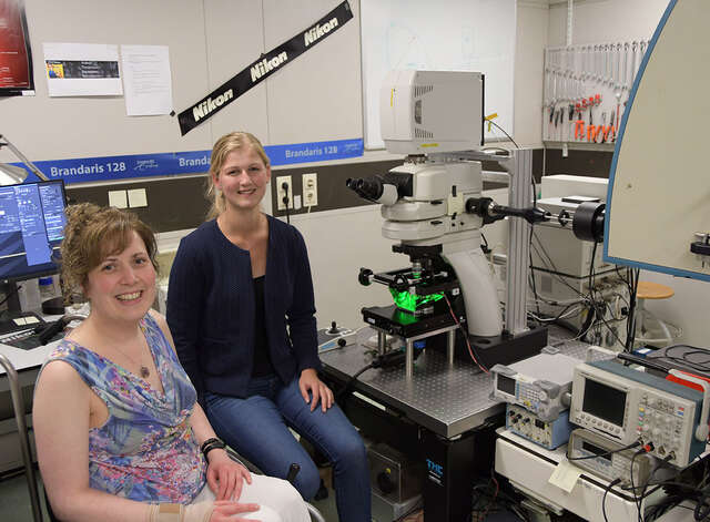
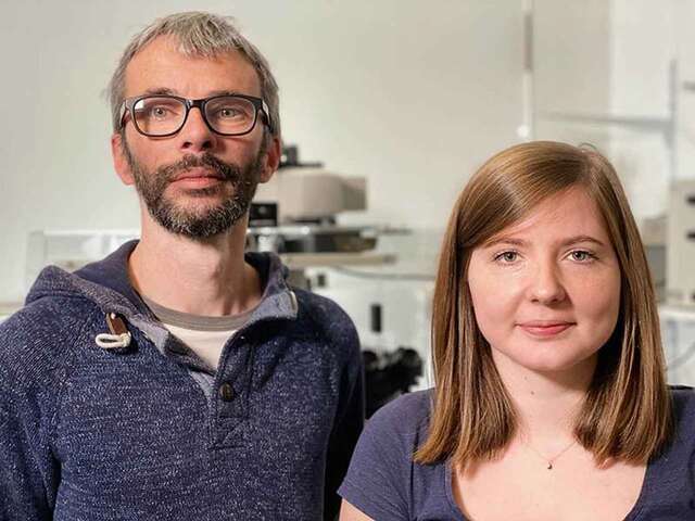
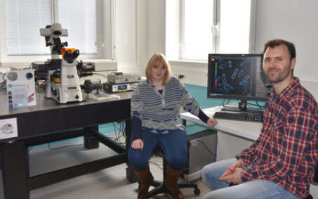
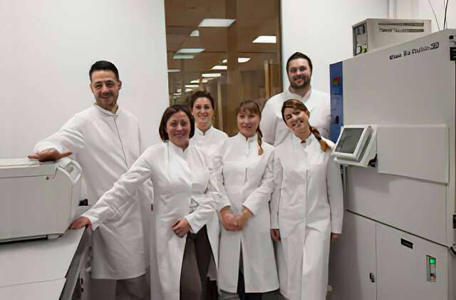
Associate Prof. Dr. Klazina Kooiman and Dr. Ines Beekers
Associate Prof. Dr. Klazina Kooiman, Head of Therapeutic Ultrasound Contrast Agent Group, and Dr. Ines Beekers, Postdoctoral Researcher in the Department of Biomedical Engineering of the Thoraxcenter, Erasmus MC, Rotterdam, the Netherlands

고객 인터뷰 - Reproduction Clinic Tokyo
“Finding the best sperm among the millions. For embryologists, this is the unique work of selection, and we feel a great sense of responsibility.”
Reproduction Clinic Tokyo, a fertility clinic located on the third floor of Shiodome City Center, a state-of-the-art office building in the Shiodome area of central Tokyo, has a large number of patients waiting for treatment, even in the evening on weekdays. We spoke with Shimpei Mizuta and Tomohiro Maekawa, who are using Nikon’s Ti2 inverted microscope for sperm sorting and Intracytoplasmic Sperm Injection (ICSI), about their thoughts on their work and their impressions of the Ti2.
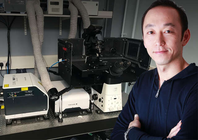
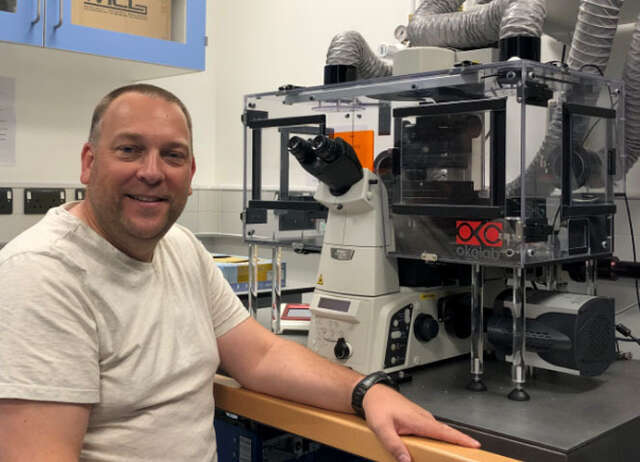
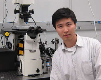
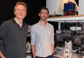
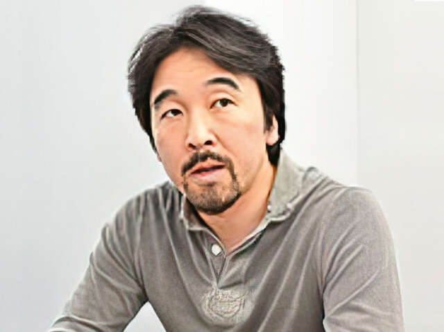
Dr. Yohei Yamauchi
Dr. Yohei Yamauchi, Principal Investigator, Cell biologist of viral infections, School of Cellular and Molecular Medicine, University of Bristol, UK
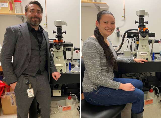
Assistant Prof. Joseph Michael Hyser & Dr. Alexandra Leigh Chang-Graham
Virology and Microbiology
Baylor College of Medicine
Houston, Texas, USA
Assistant Prof. Klazina Kooiman & Inés Beekers
Therapeutic Ultrasound Contrast Agent Group, Thoraxcenter, Department of Biomedical Engineering, Erasmus MC, Rotterdam
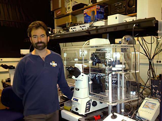
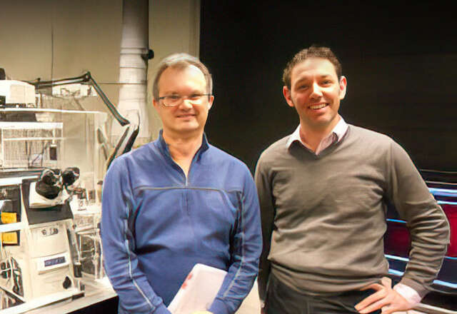
Dr. Steven Nedellec and Dr. Tiphaine Douanne
Dr. Steven Nedellec, Facility Manager of MicroPICell, Université de Nantes, France and Dr. Tiphaine Douanne, Universite de Nantes, Signaling in Oncogenesis, Angiogenesis and Permeability, CRCINA INSERM U1232, France
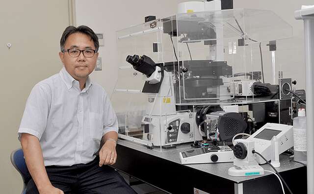
Dr. Tadahiro Iimura
Division of Bio-Imaging, Proteo-Science Center (PROS), Ehime University
Division of Analytical Bio-Medicine, Advanced Research Support Center (ADRES), Ehime University
Graduate School of Medicine, Ehime University
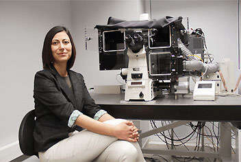
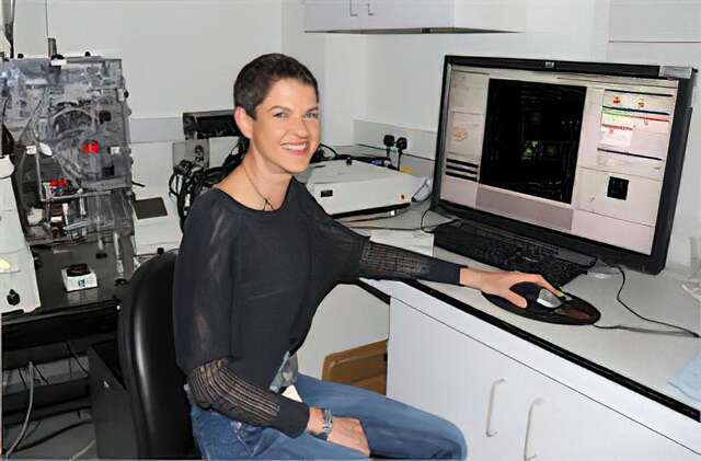
Melike Lakadamyali, Ph.D.
The advanced Fluorescence Imaging and Biophysics Group, ICFO-Institute of Photonic Sciences
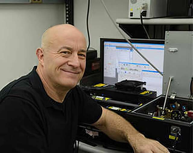
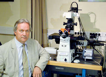
Simon C. Watkins, Ph.D.
Professor and Vice Chairman of the Dept. of Cell Biology
Director and Founder of the Center for Biologic Imaging
University of Pittsburgh
Pittsburgh, Pennsylvania, USA
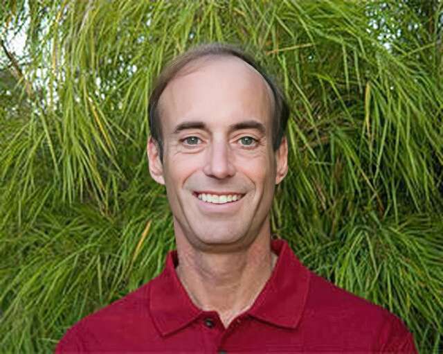
Ronald D. Vale, Ph.D.
Professor and Vice-Chairman of the Department of Cellular and Molecular Pharmacology
Investigator, Howard Hughes Medical Institute (HHMI)
The University of California, San Francisco San Francisco, CA, USA
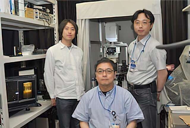
Tomomi Nemoto, Ph.D. and Ryosuke Kawakami, Ph.D.
Research Institute for Electronic Science
Sapporo, Hokkaido, Japan
Tamas Freund, Ph.D. and Istvan Katona, Ph.D.
Institute of Experimental Medicine of the Hungarian Academy of Sciences (IEM HAS)
Budapest, Hungary
Maddy Parsons, Ph.D.
Group Leader (Royal Society University Research Fellow)
Randall Division of Cell and Molecular Biophysics
King’s College London
London, United Kingdom
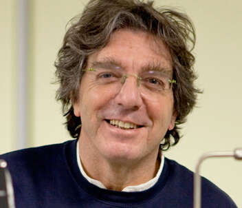
Alberto Diaspro, Ph.D.
Professor of Applied Physics, Department of Physics, University of Genoa
Director of the Department of Nanophysics, Istituto Italiano di Tecnologia
Paul Ronald Selvin, Ph.D.
Professor, Department of Physics, University of Illinois at Urbana-Champaign
Bo Huang, Ph.D.
Assistant Professor, Department of Pharmaceutical Chemistry, Department of Biochemistry and Biophysics, University of California, San Francisco
Atsushi Miyawaki, M.D., Ph.D.
Senior Team Leader, Laboratory for Cell Function Dynamics, RIKEN Brain Science Institute
Ikuo Wada, Ph.D.
Department of Cell Science, Institute of Biomedical Science, Fukushima Medical University
Romain Le Bars, Ph.D.
The Imagerie-Gif light microscopy core facility is a member of the France Bioimaging Infrastructure. The facility is hosted by the Institute for Integrative Biology of the Cell (I2BC) at Gif sur Yvette, France.
Prof. Staffan Strömblad, Ph.D.
Staffan Strömblad Ph.D. is group leader at the prestigious Karolinska Institutet, Stockholm, Sweden, an institution that awards the Physiology Nobel Prize yearly. He is also the head of the Live Cell Imaging Facility (LCI), in which the Nikon Center of Excellence for live cell imaging is integrate.
Dr. Arne Seitz, Dr. Romain Guiet and Thierry Laroche
The Faculty of Life Science (SV) at the École Polytechnique Fédéral de Laussane (EPFL), Switzerland, has a long record of excellence in research applied to life sciences
Masato Nakagawa
So-called iPS cells are attracting considerable interest as pluripotent stem cells that may open up a whole new world of medicine. The Center for iPS Cell Research and Application (CiRA) at Kyoto University is pursuing a wide range of research activities that aim to realize regenerative medicine utilizing iPS cells. The Nikon BioStation CT cell culture observation system is being used in this iPS cell research and is contributing to its efficiency.
We were pleased to have had an opportunity to speak with Masato Nakagawa, who is engaged in iPS cell research at CiRA.
Prof. Heinz Beck, Ph.D.
Full time professor at the Laboratory of Experimental Epileptology and Cognition Research hosted in the Life & Brain Center, Part of the University of Bonn.
Note: The institutions and job titles listed with each researcher reflect their affiliation at the time of the interview.