Nikon Europe B.V. | Europe & Africa
- it Change Region
- Global Site
Glial cell surrounded by axons in a rat neuronal culture labeled for microtubules and actin
Dr. Christophe Leterrier, NeuroCyto, INP, Marseille, France
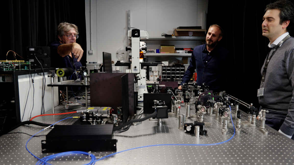
The origin of NSPARC is in the IIT (Italian Institute of Technology). We interviewed Professor Alberto Diaspro, Dr. Paolo Bianchini, and Dr. Giuseppe Vicidomini, who worked on research and development at the Nikon Imaging Center (NIC@IIT) and the Molecular Microscopy and Spectroscopy (MMS) Lab in the IIT.
Istituto Italiano di Tecnologia (IIT)
Nanoscopy
Research Team Director.
Full Professor,
Department of Physics
University of Genoa
Professor Alberto Diaspro
Prof. Diaspro: My relationship with Nikon started with the confocal laser microscope PCM 2000. This was one of Nikon’s first confocal laser microscopes and delivered great quality in terms of optics. I was developing the first Italian two-photon confocal laser microscopes at the time, and I was looking for a laser scanning microscope that was perfect for it. Nikon’s PCM2000 was ideal. This started my relationship with Nikon, which continues to this day. My current group conducts research and development for super-resolution imaging, and for NSPARC, we worked on improving the confocal laser microscope and super-resolution microscope at the same time by introducing original solutions.
Istituto Italiano di Tecnologia (IIT)
Nikon Imaging Center, NIC@IIT
Scientific Manager
Dr. Paolo Bianchini
Dr. Bianchini: In recent years, scientists who use microscopes have needs beyond simply acquiring observation images; they also want to obtain quantitative data and achieve higher spatial resolution from living biological samples. The confocal laser microscope is the type of microscope most commonly utilized in life sciences, so integrating the latest super-resolution technology, without compromising its advantages and flexibility would respond to those requirements. This was the starting point of development.
Istituto Italiano di Tecnologia (IIT)
Molecular Microscopy and Spectroscopy Lab
Principal Investigator
Dr. Giuseppe Vicidomini
Dr. Vicidomini: In 2016, when I established the Molecular Microscopy and Spectroscopy lab at the IIT, I believed that conventional confocal laser scanning microscopy is affected by a fundamental limitation. Much spatial and temporal information about the sample, potentially encoded in the image of the illumination spot, is cancelled out when using a single-element detector. To solve this limitation, the lab aimed to develop a detector array that could sample the illumination spot at the highest spatiotemporal resolution. We achieved this with the Politecnico di Milano by developing the first asynchronous-readout single-photon-avalanche-diode (SPAD) detector array. This technology crystallized to become the basis of NSPARC. We chose Nikon as a partner to expand upon these research results and transform this advance into an actual product.
Prof. Diaspro: I research photon detection and have engaged in this theme for many years alongside Dr. Vicidomini and Dr. Bianchini. For this development, I worked to assess and design the most effective method of collecting photons from samples. I was also responsible for selecting the best optical systems from the global market, such as the objective lens and the laser that would serve as a light source.
Dr. Bianchini: I believe that all of us were involved in everything from the stage of ideas to the development. I especially focused on implementing the detector array in the Nikon confocal microscope. I also carried out trial and error research to discover what level of performance could be attained through using that method. Even now, we continue searching for better ways to improve the latest confocal laser microscope performance for different samples embedded with diverse information, from single proteins to cells to tissue.
Dr. Vicidomini: Clearly, the developed detector array would serve as a core element. Still, since we were conducting joint development with Nikon, we also wanted to be meticulous about the image integration software. Together with Dr. Bianchini, Prof. Diaspro, and other researchers at IIT, we developed, and continuously updated, software designed explicitly for leveraging the novel information provided by the detector array. Eventually, we completed software that provided the high resolution we ultimately aimed for, plus a high S/N ratio. The software fully integrates with all other essential confocal features, thus allowing the maintenance of the performance of Nikon's confocal laser microscope. No compromise is introduced.
Dr. Bianchini: Spatial resolution, scanning speed, and photon budget were pointed out as issues for more advanced live-cell imaging employing confocal laser microscopes. But NSPARC enables the acquisition of much more information from samples than previously, with faster and higher-resolution imaging. As well, this system can be added to all the other confocal laser microscope system. So basically, there is no need to replace your current microscope system, making it possible to implement at a lower cost. Simplicity, flexibility, and low cost can probably be included among its greatest benefits.
Dr. Vicidomini: The NSPARC software is easy-to-use. It automatically estimates all parameters requested for the best image integration. The beauty of the NSPARC system is that it is just a confocal microscope. A novel detector and a light computation allow for gentle and flexible super-resolution imaging of your samples. Even a non-expert user employing NSPARC for the first time can operate it like the confocal laser microscope they had been utilizing before. There is no need either for special training or special sample preparation. An aspect other than simplicity is that it can image thick samples deeper and with higher resolution. NSPARC can capture detailed images of living cells inside a tissue rather than in a simple 2D cell culture. This is truly revolutionary.
Prof. Diaspro: What Dr. Vicidomini says is absolutely right. I think that to understand what is really going on in a living cell, we need information that is gained by fluorescence imaging that functions even within living cells, not just single photon detection. NSPARC makes it possible to acquire the detailed information we seek. I believe that it has the potential to bring about great advantages in the medical field as well. By utilizing the features of this system, we could design fluorescence imaging flow exclusively for cancer cells, for example. I believe that this could be the key that enables medical doctors to make critical clinical decisions in real time during surgery, as a current perspective.
Dr. Bianchini: A user asked us “How can we keep the microscope system that we have been using until now while implementing the latest confocal laser microscopy?” I believe that NSPARC, which was developed together with Nikon, is an excellent solution to that concern. I hope this new system will spread to many facilities worldwide because we believe this is indeed the next generation of confocal laser microscopy.
Dr. Vicidomini: We want to continue collaborating with Nikon to take confocal laser scanning microscopes and NSPARC to the next level. This detector array opened many new perspectives because it enabled the acquisition of enormous amounts of information from a sample, which is impossible with conventional microscopes. Indeed, this detector allows for a photon-resolved measurement, where the sample's fluorescence light is registered photon-by-photon with spatial and temporal information. Thanks to this novel single-photon microscopy paradigm, we are about to revolutionize fluorescence microscopes.
Prof. Diaspro: I think the application of artificial intelligence (AI) algorithms is approaching. It is expected that in the near future, an era of microscopy systems that comprehensively process diverse information will dawn. For this reason, we believe that there will be a need for a function where microscope imaging data that has been detected under various conditions is integrated and assessed by AI. Applying AI to a system with a confocal laser scanning microscope and NSPARC as its core will make it possible to integrate imaging data acquired from a phase-contrast microscope and differential interference microscope into fluorescent image data: label-free and fluorescence together as a powerful engine.
For this project, I was lucky to work with an excellent group of young and senior researchers here in Italy. I am more than lucky to have a cooperative relationship with Nikon R&D engineers on top of that. I hope that diverse ideas that transcend borders will continue to generate synergy.