Nikon Europe B.V. | Europe & Africa
- it Change Region
- Global Site
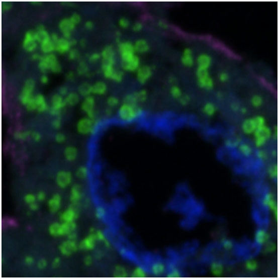
Glial cell surrounded by axons in a rat neuronal culture labeled for microtubules and actin
Dr. Christophe Leterrier, NeuroCyto, INP, Marseille, France
The newly developed Nikon Spatial Array Confocal (NSPARC) detector utilizes an ultra-low noise detector array to collect a two-dimensional image at each scanned point. This method of image scanning microscopy (ISM) improves signal-to-noise ratio by increasing the available signal level while simultaneously allowing imaging with lower excitation power.
Single-photon sensitivity and array detection extend the capabilities of the AX system by revealing unseen details in every image, with array detection pushes the boundaries of resolution beyond the theoretical limits.
NSPARC’s array detection provides spatial sampling of the confocal point spread function in every pixel. This method detects information that can be utilized to extract enhanced resolution performance both laterally and axially. Built-in variable emission optics increase flexibility to allow users to replicate the function of a pinhole. This means the ability to prioritize enhanced optical sectioning or increased overall signal collection depending upon specific experimental needs.
The variable emission optics allow Nikon to offer many compatible low and high mag objectives to match specimens. This includes silicon immersion objectives which minimize refractive index mismatches, improving overall system performance.
Coupled with the AX/AX R's ultra-large 25mm FOV, the system supports a wide selection of objectives capable of obtaining image data from large overviews down to extremely fine details, which can then be measured and analyzed.
An extremely low noise profile along with incredible sensitivity renders outstanding results even under demanding imaging conditions, including high speed resonant imaging.
As an added benefit, additional spatial information is acquired for every pixel using the detector array, which can be used to further enhance resolution and brightness.
NSPARC Noise Profile
GaAsP Noise Profile
Comparing NSPARC and GaAsP bias images (acquisition with no light to measure noise) reveal extremely low standard deviation in pixel-to-pixel intensity, significantly lower than GaAsP or PMT detectors.
NSPARC
Traditional Detector
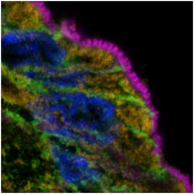
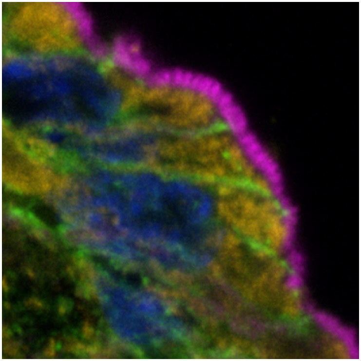
The extremely low noise of NSPARC detectors and the additional spatial data collection per pixel result in high signal-to-noise, sharper images compared to standard GaAsP or PMT images.
The combination of ultra-short dwell times with the AX R resonant scanner and NSPARC’s sensitivity allows longer, less phototoxic imaging data to be collected in live-cell assays.
Live cell imaging timelapse using AX resonant scanning of mitochondrial dynamics using Plan Apo Lambda 100x objective lens (an area of the large FOV timelapse is zoomed here to show every 10th frame of a fast acquisition)
With single photon detection sensitivity, the NSPARC detector’s extremely low noise profile and exceptional sensitivity make it a perfect match for AX R confocal resonant high-speed imaging, where the pixel dwell time is as short as 200 nanoseconds, enabling image acquisition at video frame rate. More details can be extracted from images for downstream analysis and computation.
Video-rate timelapse imaging with AX R of zebrafish vasculature was performed using NSPARC and conventional detectors to compare their sensitivities. Imaging kymographs were created (yellow lines) across a vessel in order to visualize the vessel endothelium position over time. Because of its array detection and increased SNR, the NSPARC kymograph more clearly displays the pulsatory nature of the endothelial layer over time, in comparison to a conventional GaAsP detector, where these are not resolved.
Where a point scanning detector traditionally produces an intensity output only for each pixel, NSPARC comprises an array of 25 detectors that operates more like an extremely sensitive camera: two-dimensional spatial information from each scanned pixel is collected by the detector.
Optical lenses direct emission light to the detector, allowing it to be used with various objectives and magnifications, while simultaneously allowing the user to define the size of the illumination spot on the detector array. This enables oversampling of the conventional single airy unit emission from the confocal plane.
This two-dimensional information is immediately used to obtain ultra-fine structural information, which is lost in conventional detection.
Each pixel from NSPARC contains spatial information, which can be used to reconstruct fine details in a result image.
With NSPARC detection, the fluorescence emission light is directed through optics to the array detector, where the projected light fills the array.
Each pixel from a traditional detector contains only one intensity value, and no spatial information.
With traditional PMT or GaAsP detectors, the fluorescence emission light passes through a variable sized emission pinhole (typically set to 1 airy unit).
When the NSPARC detector is combined with the AX R resonant scanner, super-resolution images of large areas of 2048 x 2048 pixels can be acquired at high speed (approximately 6 times* faster than with a galvano scanner). It is capable of capturing rapid changes in minute structures over large areas.
* When using the CFI Plan Apochromat Lambda D 60X Oil objective and a 2.5X zoom.
For the same pixel size, a 2K x 2K pixel image provides four times the area of a 1K x 1K pixel image.
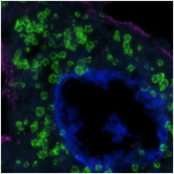
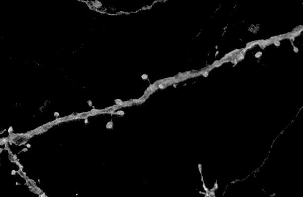
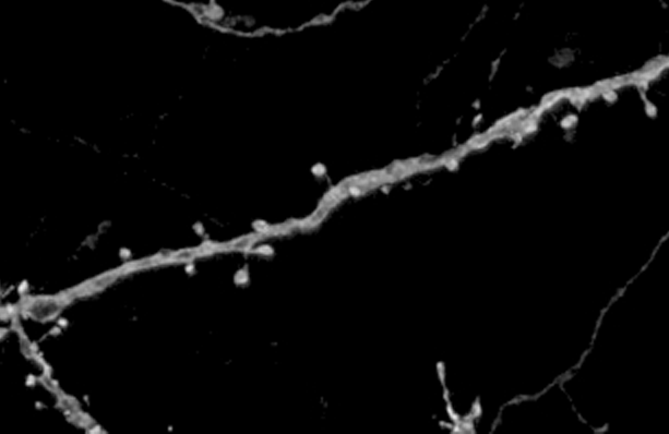
Thy1-EGFP mouse neuron (after tissue clearing)
Sample courtesy of: Lin Daniel, PhD. SunJin Lab Co.
For the same imaging area, a 2K x 2K pixel image provides higher image resolution than a 1K x 1K pixel image.