- it Change Region
- Global Site
- Casa
- Prodotti
- Microscopi confocali e multifotonici
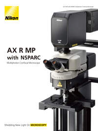
- AX R MP with NSPARC
Robust multiphoton system tailored for in vivo applications
Nikon’s multiphoton confocal microscopes, which clearly visualize fine structures deep within living organisms, have evolved even further. The AX R MP is equipped with a high-speed resonant scanner with 2K resolution and can capture in a single scan dynamics that span a wide area with superior spatial and temporal resolution.
In addition, the innovative NSPARC super-resolution detector utilizes a newly developed SPPC array detector to collect a two-dimensional image at each scanned point, achieving a significant improvement in resolution. This enables macro-to-micro imaging with a single microscope system.
The AX R MP maximizes image size, as well as spatial and temporal resolution, providing a uniquely powerful solution for any multiphoton experiment.
Caratteristiche principali
Bigger pictures. Fast.
Featuring a a field-of-view of FN22 for both resonant and galvano scanners, the AX R MP captures more data in every frame at any magnification. This means less time spent imaging large specimens and more information in every frame.
See more at high magnification
Use the AX R MP field of view and Nikon's world-class objective lenses to capture large specimens in one image. With resolutions up to 8K x 8K, these images can be captured without sacrificing a single detail.
Redesigned optics maximize performance over a large area and deliver seamless image stitching at much higher speeds. These improvements save time during acquisition and minimize post-processing.
SHG image of mouse jaw, Vessel: Alexa 488
Z-stack image acquired from a 12 x 5 stitched image in 6.7 x 16.2 mm area. Image courtesy of Lin Daniel, PhD, SunJin Lab Co.
Objective: CFI Plan Apochromat Lambda D 10
Embryonic Zebrafish Blood Vessels (0:10)
Video of the 3D distribution of blood vessels in a 700 µm thick Z stack acquired in an embryonic zebrafish labeled with GFP and RFP.
Acquired with a CFI75 Apochromat LWD 20XC W objective lens.
Courtesy Erika Dreikorn and Dr. Beth Roman, Department of Human Genetics, University of Pittsburgh Graduate School of Public Health
High-speed imaging that captures the dynamics of life
The 2K x 2K resonant scanner featured on the AX R MP provides fast (up to 720 fps), gentle and high resolution imaging. This means samples live longer and stay healthier all while much higher-quality data is being collected. Alternatively, the resonant scanner can be applied to large fixed samples to dramatically improve the speed of large volume acquistions, with no loss of image integrity.
Embryonic zebrafish, Blood vessels: GFP, Blood cell: RFP (0:10)
Individual blood cells are identified in high resolution, and blood flow is imaged at a high speed of 28 fps (2048 x 546 pixels)
Images courtesy of Erika Dreikorn and Dr. Beth Roman, Department of Human Genetics, University of Pittsburgh Graduate School of Public Health
Objective: CFI75 Apochromat LWD 20XC W
Bright, high-definition imaging of deep structures
Utilizing high-resolution resonant scanning, extremely low-noise detectors and efficient multiphoton excitation, the AX R MP collects fluorescence from deep within any specimen for bright, high-definition results.
High resolution deep imaging for intravital microscopy
The AX R MP's two selectable scanners, resonant and Galvano, allow users flexibility in acquisition, and provide both high-speed and high-resolution solutions. The Galvano scanner is capable of obtaining 8192 x 8192 pixel high resolution images, with a pixel density that enables Nyquist sampling at any magnification. The high-speed resonant scanner supports high resolution imaging with pixel densities of up to 2048 x 2048. Both can visualize morphological changes in deeper regions in fine detail.
Transgenic Mouse Cerebellum Neurons (0:04)
Cutaway video of Purkinje neurons in the cerebellum of an LC3-GFP transgenic mouse acquired with a CFI75 Apochromat LWD 20XC W objective lens and deconvolved using the NIS-Elements software.
Courtesy of Dr. L. Dubreil, Dr. J. Pichon and Pr MA Colle, CENN at PAnTher UMR703 INRAE/Oniris, Nantes France
0 μm
-150 μm
-300 μm
-450 μm
Amazingly sensitive
Replaceable DM/IR cut filter kit
The AX R MP maximizes signal collection by placing detectors as close to the sample as possible. Sensitivity is improved by utilizing the latest electrical and detector designs to drastically reduce noise that normally obscures dim signals. Detector configurations are flexible allowing the system to be tailored to any experimental needs.
Space to expand
The AX-FN series of microscopes prioritizes flexibility and free space under the objective. This means experimental design is limited only by the users’ imagination, rather than the body of the microscope.
Gate stand
For systems requiring depth
Single stand
For systems requiring breadth
The AX R MP can also be mounted on an ECLIPSE Ti2 inverted microscope.
The motorized upright microscope dedicated for AX R MP provides up to 40 cm under the objective in two unique configurations. Both stands provide a large amount of free space around the sample improving positioning, flexibility and accessibility.
Con gate stand
Con stand singolo
A system built for intravital microscopy
The AX R MP is designed from top to bottom to deliver maximum performance and flexibility for intravital imaging. Built to be fast, without sacrificing image quality, this system delivers reliable data in even the most difficult conditions. With unique AX-FN microscope stands, space is prioritized allowing access to new experimental designs and samples. Auto-alignment brings day-to-day reliability that is absolutely essential when precious intravital data is being collected.

Accommodate any sample
Experiments are best performed with minimal disturbance to the sample. Utilizing Nikon's new CFI75 tilting nosepiece, the objective can be oriented to image samples in their natural positions, minimizing stress and uncontrolled variables. This nosepiece is compatible with Piezo Z, maximizing 3D image speed in these challenging samples.
Movimento grossolano dell'asse Z (±3 mm)
Corsa del dispositivo Piezo Z: 450 μm
Rotazione verticale (±90°)
Rotazione orizzontale (±100°)
Simultaneous stimulation imaging
The AX R MP supports simultaneous photostimulation imaging with two wavelengths. In addition to versatile visible light stimulation, it is also capable of long-wavelength infrared light stimulation, which enables deep stimulation.
Opti-Microscan photostimulator (optional)
Photostimulation using wavelengths* of 400 nm, 488 nm, and 561 nm enables simultaneous visible light stimulation and IR imaging. Stimulation modes include simultaneous, sequential, and manual stimulation.
* Limited by the specifications of the filter cube used.
IR photostimulation AX-STM-IR IR stimulation unit (option)
This allows IR imaging using wavelengths of 750 - 950 nm / 1030 - 1070 nm, while stimulating the sample at IR wavelengths of 1030 - 1070 nm / 900 - 950 nm.
IR stimulation / IR imaging
In Purkinje cell dendrites of an adult mouse cerebellum, rsChrmine is activated by locally stimulating the position indicated by the arrow with a 1060 nm laser, while imaging jGCaMP7f signals at 7.5 fps using a 920 nm laser.
Image courtesy of Dr. Naofumi Uesaka, Neurophysiology, Graduate School of Medical and Dental Sciences, Institute of Science Tokyo
Support for visible light imaging
The AX R MP supports observation not only at infrared wavelengths, but also at visible wavelengths. It enables both multiphoton imaging and confocal imaging with a single microscope. It also enables simultaneous photostimulation and imaging using two different wavelengths.
Embryonic Zebrafish Blood Vessels
Blood vessels in an embryonic zebrafish labeled with GFP and RFP. Imaged using a CFI75 Apochromat LWD 20XC W objective lens.
Courtesy of Erika Dreikorn and Dr. Beth Roman, Department of Human Genetics, University of Pittsburgh Graduate School of Public Health
DUX-VB high-sensitivity visible light detector unit
The transmission wavelength band of the LVF (Linear Variable Filter) employed in the DUX-VB gradually changes depending on its location, enabling continuous tuning of the wavelength detection setting within a range of 400 nm to 750 nm. From 2 to 4 channels can be selected, and high sensitivity GaAsP PMT can be used for all channels.


