Nikon Instruments Inc. | Americas
- es Change Region
- Global Site
noviembre 2021
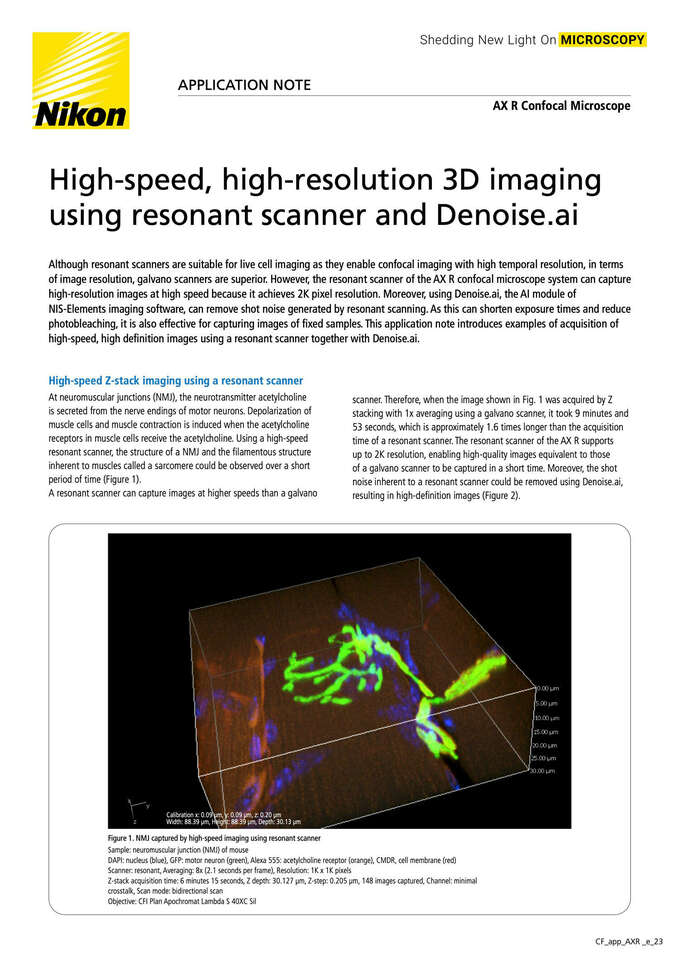
Although resonant scanners are suitable for live cell imaging as they enable confocal imaging with high temporal resolution, in terms of image resolution, galvano scanners are superior. However, the resonant scanner of the AX R confocal microscope system can capture high-resolution images at high speed because it achieves 2K pixel resolution. Moreover, using Denoise.ai, the AI module of NIS-Elements imaging software, can remove shot noise generated by resonant scanning. As this can shorten exposure times and reduce photobleaching, it is also effective for capturing images of fixed samples. This application note introduces examples of acquisition of high-speed, high definition images using a resonant scanner together with Denoise.ai.
agosto 2021
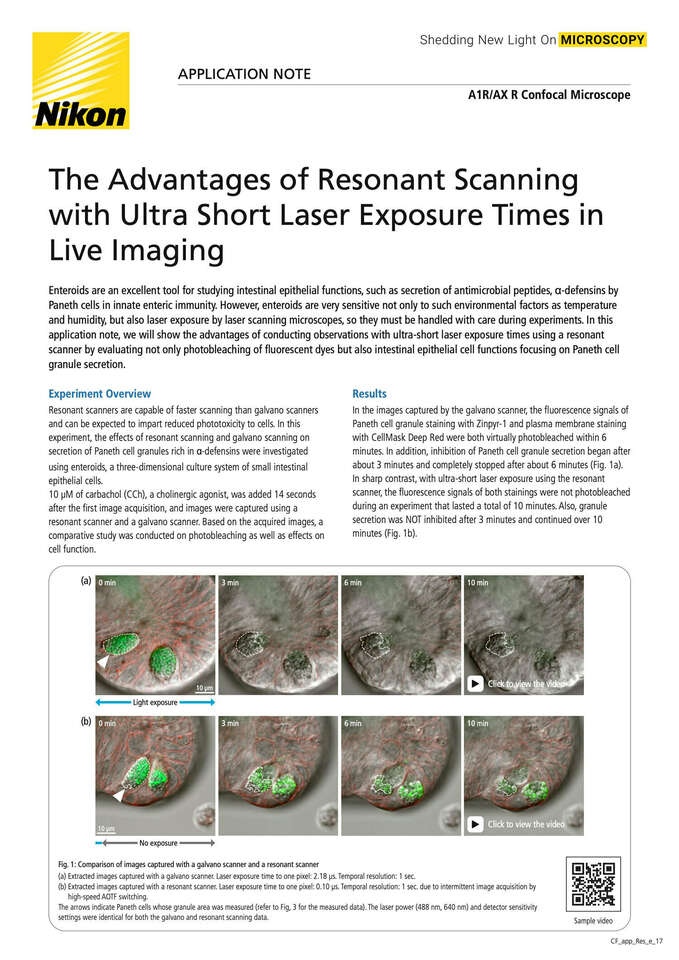
Enteroids are an excellent tool for studying intestinal epithelial functions, such as secretion of antimicrobial peptides, α-defensins by Paneth cells in innate enteric immunity. However, enteroids are very sensitive not only to such environmental factors as temperature and humidity, but also laser exposure by laser scanning microscopes, so they must be handled with care during experiments. In this application note, we will show the advantages of conducting observations with ultra-short laser exposure times using a resonant scanner by evaluating not only photobleaching of fluorescent dyes but also intestinal epithelial cell functions focusing on Paneth cell granule secretion.
noviembre 2021
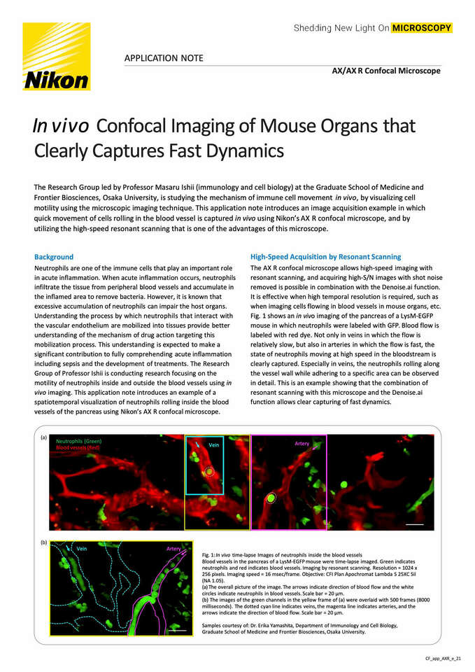
The Research Group led by Professor Masaru Ishii (immunology and cell biology) at the Graduate School of Medicine and Frontier Biosciences, Osaka University, is studying the mechanism of immune cell movement in vivo by visualizing cell motility using the microscopic imaging technique. This application note introduces an image acquisition example in which quick movement of cells rolling in the blood vessel is captured in vivo using Nikon’s AX R confocal microscope, and by utilizing the high speed resonant scanning that is one of the advantages of this microscope.
junio 2021
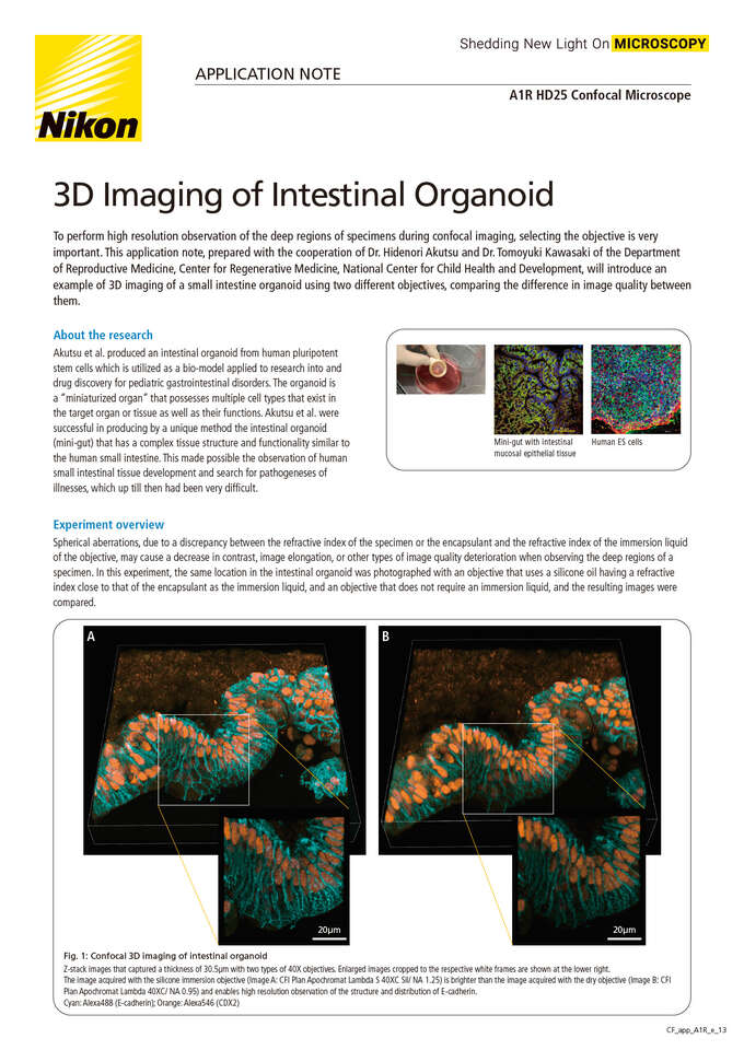
To perform high resolution observation of the deep regions of specimens during confocal imaging, selecting the objective is very important. This application note, prepared with the cooperation of Dr. Hidenori Akutsu and Dr. Tomoyuki Kawasaki of the Department of Reproductive Medicine, Center for Regenerative Medicine, National Center for Child Health and Development, will introduce an example of 3D imaging of a small intestine organoid using two different objectives, comparing the difference in image quality between them.