
Specifications
| Model | Dimensions | Transmittance | NA | W.D. (mm) | Cover glass thickness | Correction ring | Observation |
|---|---|---|---|---|---|---|---|
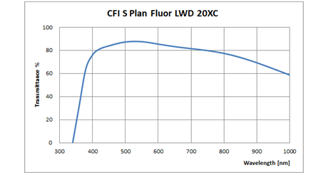
| CFI S Plan Fluor LWD 20XC | Diagram | graph | 0.70 | 1.3-2.3 | 0-1.8 | ∨ | BF, DF (Dry/Oil), DIC, POL, FL (visible light, UV) |
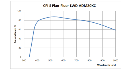
| CFI S Plan Fluor LWD ADM 20XC | Diagram | graph | 0.70 | 1.3-2.3 | 0-1.8 | ∨ | BF, DF (Dry/Oil), PH, FL (visible light, UV) |
BF: Brightfield
DF: Darkfield
PH: Phase contrast
POL: Simple polarizing
FL: Fluorescence
*Possible but not recommended
**External phase contrast observation is possible with Eclipse Ti2-E


