- fr Change Region
- Global Site
- Accueil
- Ressources
- Notes d'application
Notes d'application

Speedy and clear 3D imaging of organoids
An efficient workflow using epi epi-fluorescence and confocal imaging
mars 2025
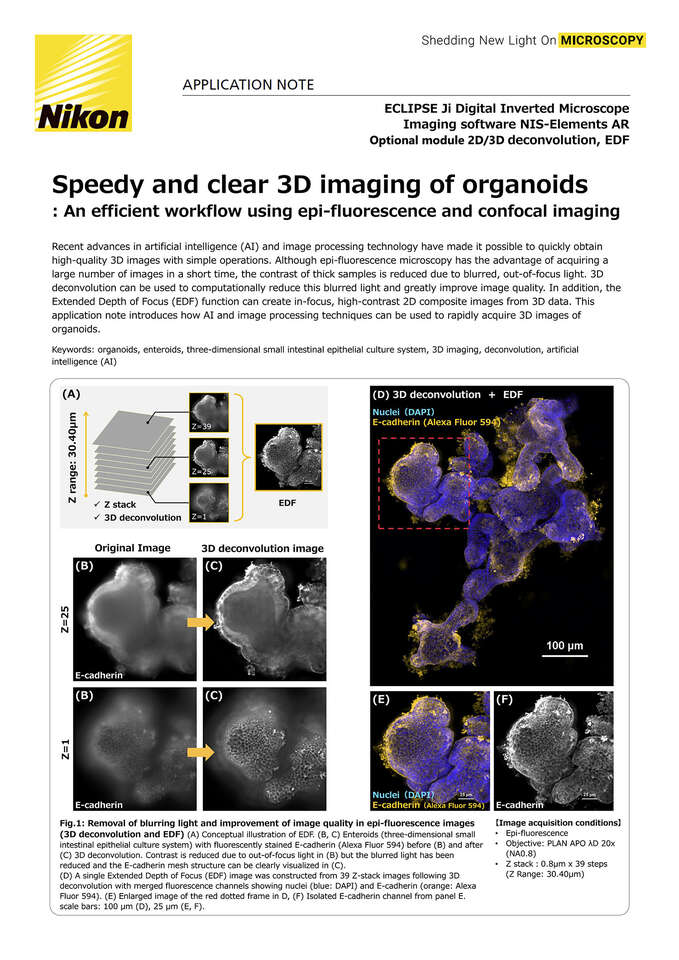
Recent advances in artificial intelligence (AI) and image processing technology have made it possible to quickly obtain high -quality 3D images with simple operations. Although epi epi-fluorescence microscopy has the advantage of acquiring a large number of images in a short time, the contrast of thick samples is reduced due to blurred, out out-of -focus light. 3D deconvolution can be used to computationally reduce this blurred light and greatly improve image quality. In addition, the Extended Depth of Focus (EDF) function can create in in-focus, high high-contrast 2D composite images from 3D data. This application note introduces how AI and image processing techniques can be used to rapidly acquire 3D images of organoids.
Keywords: organoids, enteroids, three three-dimensional small intestinal epithelial culture system, 3D imaging, deconvolution, artificial intelligence (AI)
- Accueil
- Ressources
- Notes d'application
