Nikon Europe B.V. | Europe & Africa
- fr Change Region
- Global Site
avril 2023
Mixed connective tissue and epithelial tumors of the breast mainly include fibroadenoma (FA) and phyllodes tumor (PT). Fibroadenoma is the most common breast tumor in young women, while phyllodes tumor is a relatively rare disease, accounting for about 0.5% of all breast tumors. Whereas fibroadenomas are usually 2-3 cm in size and stop growing, phyllodes tumors grow rapidly and can grow into massive tumors exceeding 10 cm. Also, phyllodes tumors are benign tumors like fibroadenomas, but they may become malignant during the process of growth.
Fibroadenomas and phyllodes tumors are often difficult to distinguish by needle biopsy because of their similarities in clinical and histological images, despite differences in surgical indications and surgical techniques. Several studies have been conducted in the past to search for factors useful in differentiating between fibroadenoma and phyllodes tumors, but none have yet been established.
Therefore, Dr. Kana Kobayashi-Taguchi and Dr. Yoshiaki Kamei of the Breast Center, Ehime University Hospital, and Dr. Takashi Saitou and Dr. Takeshi Imamura of the Department of Molecular Medicine for Pathogenesis, Graduate School of Medicine, Ehime University, attempted image diagnosis of breast tumors using multiphoton microscopy. This application note introduces the contribution of multiphoton microscopes to medicine, based on the research results of Dr. Taguchi et al.
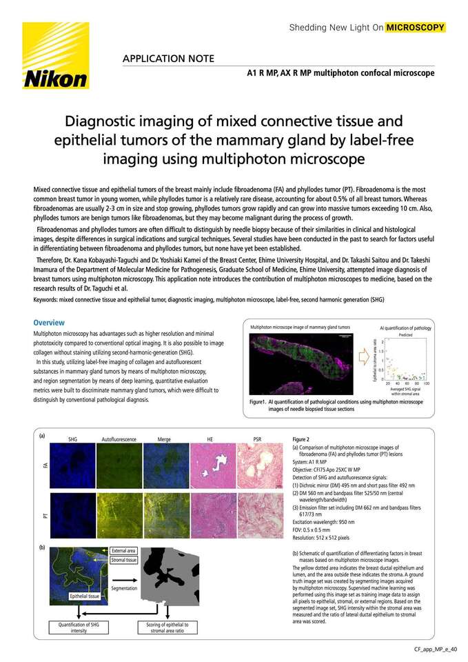
Figure 1. AI quantification of pathological conditions using multiphoton microscope images of needle biopsied tissue sections
Multiphoton microscopy has advantages such as higher resolution and minimal phototoxicity compared to conventional optical imaging. It is also possible to image collagen without staining utilizing second-harmonic-generation (SHG).
In this study, utilizing label-free imaging of collagen and autofluorescent substances in mammary gland tumors by means of multiphoton microscopy, and region segmentation by means of deep learning, quantitative evaluation metrics were built to discriminate mammary gland tumors, which were difficult to distinguish by conventional pathological diagnosis.
Figure 2
(a) Comparison of multiphoton microscope images of fibroadenoma (FA) and phyllodes tumor (PT) lesions
System: A1 R MP
Objective: CFI75 Apo 25XC W MP
Detection of SHG and autofluorescence signals:
(1) Dichroic mirror (DM) 495 nm and short pass filter 492 nm
(2) DM 560 nm and bandpass filter 525/50 nm (central wavelength/bandwidth)
(3) Emission filter set including DM 662 nm and bandpass filters 617/73 nm
Excitation wavelength: 950 nm FOV: 0.5 x 0.5 mm
Resolution: 512 x 512 pixels
(b) Schematic of quantification of differentiating factors in breast masses based on multiphoton microscope images.
The yellow dotted area indicates the breast ductal epithelium and lumen, and the area outside these indicates the stroma. A ground truth image set was created by segmenting images acquired by multiphoton microscopy. Supervised machine learning was performed using this image set as training image data to assign all pixels to epithelial, stromal, or external regions. Based on the segmented image set, SHG intensity within the stromal area was measured and the ratio of lateral ductal epithelium to stromal area was scored.
Figure 3. Deep learning using multiphoton microscope images
(a)Results of segmentation in the training image set. From the left, the original multiphoton microscope images, ground truth images, predicted images, and difference images. Differences between images show the FN regions in magenta and FP regions in green.
(b)Results of segmentation on the test image set.
(c)Numerical evaluation of segmentation results. Total accuracy between the ground truth and predicted images and the weighted IoU, which is the area-weighted sum of the IoU values for each image, are shown.
Figure 4. Establishment of quantitative discriminative factors for breast masses using multiphoton microscope images
(a)Epithelial to stromal area ratio of FA and PT lesions
(b)Mean SHG signal intensity within the stromal area of FA and PT lesions
(c)Scatterplot with the two quantification scores. Solid and hollow circles respectively indicate FA and PT data. Circles of the same color represent samples obtained from the same patient.
Comparing the HE- and PSR-stained images with the SHG images acquired by multiphoton microscopy, the SHG images showed dark ductal epithelium and strong signals in the collagen-rich stromal regions. Collagen observed in SHG images resembled in shape and pattern the fibril structures in PSR-stained images that specifically stained type I and type III collagen. In addition, the boundary between epithelium and stroma was confirmed in the autofluorescence image obtained by multiphoton microscopy shown in green (Fig. 2 (a)).
Mixed connective tissue and epithelial tumors are lesions with both epithelial and stromal proliferation, but phyllodes tumor (PT) has been reported to have more pronounced stromal proliferation than fibroadenoma (FA). To establish quantitative criteria to distinguish between FA and PT, scoring of the area ratio of epithelium and stroma was attempted using image segmentation by means of deep learning (Fig. 2(b), Fig. 3). Ground truth images were prepared from multiphoton microscope images in which three regions, epithelium, stroma, and outer, were manually labeled based on HE-stained images. The overall accuracy and the intersection of union (IoU) between the predicted and ground truth images were compared, and the results showed that the test image set had a total accuracy of 93.5% and an IoU of 89.5%, indicating high segmentation performance (Figure 3(c)).
The standard deviation was then calculated for the images for which epithelial-to-stromal area ratio scoring was performed based on the results of the image segmentation analysis. As a result, PT showed a higher deviation than FA in both ground truth and predicted image data (Fig. 4(a)). In addition, quantification of the SHG signal intensity in the stromal regions showed a stronger SHG signal in FA than in PT (Fig. 4 (b)). When a scatter plot combining the score of the epithelial-stromal area ratio and the SHG signal intensity of the stromal area was created, FA and PT could be clearly separated (Fig.4 (c)).
In multiphoton microscope images, the ductal epithelial regions exhibiting autofluorescence and the stromal regions exhibiting collagen-derived SHG signals were segmented using SegNet, an image segmentation program utilizing deep learning. When quantifying the ductal epithelial/stromal area ratio and the signal intensity of SHG within the stromal area, the former was higher in PT and the latter in FA. When an attempt was made to distinguish between PT and FA using these two factors, a clear distinction was attainable.
Autofluorescence imaging combined with multiphoton microscopy and AI image analysis identified quantitative factors that enabled the differential diagnosis of fibroadenoma and phyllodes tumor. It is expected that this result will lead to this method’s application in computer-aided diagnosis of mixed connective tissue and epithelial tumors in mammary glands.
“Computer-Aided Detection of Quantitative Signatures for Breast Fibroepithelial Tumors Using Label-Free Multi-Photon Imaging”
Kana Kobayashi-Taguchi, Takashi Saitou, Yoshiaki Kamei, Akari Murakami, Kanako Nishiyama, Reina Aoki, Erina Kusakabe, Haruna Noda, Michiko Yamashita, Riko Kitazawa, Takeshi Imamura, Yasutsugu Takada
Molecules. 2022 May 23;27(10):3340. doi: 10.3390/molecules27103340.
Department of Breast Center, Ehime University Hospital:
https://www.m.ehime-u.ac.jp/hospital/breast/?page_id=215
Department of Molecular Medicine for Pathogenesis, Graduate School of Medicine, Ehime University:
https://www.m.ehime-u.ac.jp/school/imaging/
AX R MP Multiphoton Confocal Microscope


