- es Change Region
- Global Site
- Casa
- Productos
- Microscopios confocales y multifotónicos
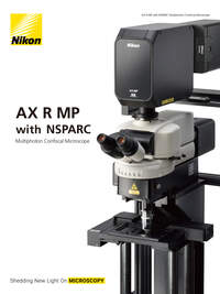
- AX R MP with NSPARC
AX R MP with NSPARC
Sistema de microscopio multifotónico
Especificaciones
Main body | AX-FNSP | AX-FNGP |
|---|---|---|
Optical system | Infinity optical system | |
Microscope stands | AX-FNSP Single Stand | AX-FNGP Gate Stand |
Focusing | AX-FN Focusing Nosepiece Unit | |
Controls | AX-FNCTL Control Box | |
Stages | AX-FNSP | AX-FNGP |
|---|---|---|
Adapter | AX-FNSA Stage Adapter, supporting both manual and motorized XY stages. Stage height: adjustable to 2 positions depending on sample size (400 mm/200 mm from the surface of the vibration isolated table) | |
Stage | FN-3PS2 XY stage, Cross travel 29.5 (X) x 29.5 (Y) mm, with 2 auxiliary plates | |
Epi-fluorescent illuminator | AX-FNSP | AX-FNGP |
|---|---|---|
Illumination unit | NI-FLEI-2 Epi-fluorescence attachment | |
Light source | D-LEDI Fluorescent LED Illumination System | |
Filter cube turret | 6 mountable filter cubes, shutter function
| |
Photostimulation device | AX-FNBPU Stimulation Back Port, 6 mountable filter cubes, Fluorescence imaging and simultaneous stimulation imaging can be switched | |
Diascopic illuminator | AX-FNSP | AX-FNGP |
|---|---|---|
Illumination unit | AX-FNDIA Diascopic Unit | — |
Light source | C-LL High Color Rendering LED Lamphouse | — |
Shutter | NI-SH-E Motorized Shutter | — |
Condenser | FN-C LWD condenser, O.D. 8.2 mm, NA: 0.78 | — |
Polarizer Turret | NI-PT Polarizer Turret, Visible or infrared polarizer attachable | — |
AX-FNSP | AX-FNGP | |
|---|---|---|
Tubes | Pupillary distance: 50-75 mm, Inclination angle: 15-35 degrees, Eyepiece/Upper port/Rear port: 100/0/0, 0/100/0, 0/0/100 via DSC Zooming Port
| |
Eyepieces (FN) | CFI 10X (22), CFI 12.5X (16), CFI 15X (14.5), CFI UW 10X (25) | |
Photodetector | AX-NEU Non-descanned EPI Upright Detector | |
Nosepieces |
| |
Observation methods | Brightfield, Epi-fluorescence, DIC | |
Power consumption | 100W | |
Weight (approx.) | 66 kg (fully motorized fluorescence system, with diascopic illuminator) | 66 kg (fully motorized fluorescence system) |
Scan head | AX R MP |
|---|---|
Type | AX-SHRM AX R MP Scan Head & Controller |
Field number (FN) | φ22 mm |
Standard image acquisition | Galvano scanner |
High-speed image acquisition | Resonant scanner |
Scan mode | Line scanning, bi-direction scanning and averaging |
Simultaneous acquisition | Max. 5 channels (including a diascopic detector channel) |
IR laser wavelength range | 700-1080 nm (1080 system), 820-1300 nm (1300 system) |
Dichroic mirror | Position: 6 |
Pinhole | 6-153 µm variable |
Zoom | 1–1000X continuously variable |
Input/output port | 2 laser input ports |
Laser for multiphoton microscopy | AX R MP |
|---|---|
Single 1080 system | Mai Tai HP/eHP DeepSee, Chameleon Vision II, Axon 920*6,Chameleon Discovery LX |
Dual 1080 system | Chameleon Vision II + Axon 920*6, Axon 920*6 + Axon 1064*7, Chameleon Discovery LX + Axon 920*6 |
Single 1300 system | InSight X3 + InSight X3+, Chameleon Discovery NX |
Dual 1300 system | InSight X3 Dual Option, InSight X3+ Dual Option, Chameleon Discovery NX, Chameleon Discovery NX + Axon 920*6 |
Incident optics | 700-1080 nm (1080 system), 820-1300 nm (1300 system), auto alignment |
Modulation | Method: AOM (Acousto-Optic Modulator) device |
Laser for confocal microscopy (option) | AX R MP |
|---|---|
4-laser unit | 405 nm, 488 nm, 561 nm and 640 nm lasers are installed |
5-laser unit | 405 nm, 488 nm, 561 nm, 594 nm and 640 nm lasers are installed |
6-laser unit | 405 nm, 445 nm, 488 nm, 515 nm, 561 nm and 640 nm lasers are installed |
NDD for multiphoton microscopy | AX R MP |
|---|---|
NDD EPI unit AX-NEI (for Ti2-E) and AX-NEU (for AX-FNSP/FNGP) | Detectable wavelength range: 400-650 nm (1080 system), 400-750 nm (1300 system) |
Visible stimulation/IR imaging (option) | AX R MP |
|---|---|
Opti-Microscan (for AX-FNSP) | Stimulation wavelength: 405 nm, 488 nm, 561 nm; |
Diascopic detector (option) | AX R MP |
|---|---|
AX-DUT-MP (for AX-FNSP/Ti2-E)*8 | Detectable wavelength range: 400-920 nm |
Detector for confocal / multiphoton microscopy (option) | AX R MP |
|---|---|
DUX-VB detector unit | Detectable wavelength range: 400-650 nm (with IR laser), 400-750 nm (with visible laser); Detection width: 10 nm to 320 nm |
DUX-ST detector unit*9 | Detectable wavelength range: 400-750 nm |
NSPARC Detector Unit | Equipped with SPPC (Single Pixel Photon Counter) array detector |
Option | AX R MP |
|---|---|
Motorized XYZ | Motorized XY stage (for AX-FNSP/FNGP/Ti2-E), High-speed piezo Z stage (for Ti2-E), High-speed piezo objective-positioning system (for AX-FNSP/FNGP) |
Nosepiece for AX-FNSP/FNGP | AX-FNTN-H CFI75 single tilting nosepiece*4 |
Software | AX R MP |
|---|---|
Acquisition/analysis | Imaging software (equipped with Denoise.ai noise reduction function): NIS-Elements C or NIS-Elements C-ER |
Display/image generation | 2D analysis, 3D volume rendering/orthogonal, 4D analysis, spectral unmixing |
Image format | JP2, JPG, TIFF, BMP, GIF, PNG, ND2, JFF, JTF, AVI, ICS/IDS |
Application | FRAP, FLIP, FRET(option), photoactivation, 3D time-lapse imaging, multipoint time-lapse imaging, colocalization |
Control computer | AX R MP |
|---|---|
OS | Windows®10 Pro 64 bit, Microsoft Windows® 11 Pro*10 |
AX R MP | |
|---|---|
Visible stimulation/IR imaging (option) | Stimulation wavelength: 405 nm, 488 nm, 561 nm |
Compatible microscopes | Dedicated AX-FNSP/AX-GNGP motorized upright microscope system, ECLIPSE Ti2-E motorized inverted microscope |
Z step | AX-FNSP/FNGP: 0.02 µm, Ti2-E: 0.02 µm |
Recommended installation conditions | Temperature 20 - 25˚C, ± 1˚C, air conditioning at all hours |
IR stimulation/ IR Imaging (OPTION) | AX R MP |
|---|---|
AX-STM-IR IR stimulation unit*8 | Stimulation wavelength: 1030-1070 nm, 900-950 nm |
*1 Based on the focus position
*2 Software controlled value
*3 DIC prism slider cannot be attached
*4 FN12, Usable objectives: CFI75 LWD 16X W, CFI75 Apochromat LWD 20XC W, CFI75 Apochromat 25XC W, CFI75 Apochromat 25XC W 1300
*5 Cannot be used with diascopic illumination. The FN-MN-H cannot be used with diascopic illumination only when the 400 μm objective piezo positioner (PI) is attached.
*6 Axon 920 : Axon 920-1、Axon 920-2
*7 Axon 1064 : Axon 1064-1、Axon 1064-3
*8 Cannot be mounted on AX-FNGP
*9 Must be used with a confocal laser.
*10 Windows 10 and Windows 11 are trademarks or registered trademarks of Microsoft Corporation in the United States and other countries.

