니콘인스트루먼트코리아 | Korea
- ko Change Region
- Global Site
Tissue culture is the process of maintaining cells in vitro. Cells are routinely kept in suspension, adhering to the surface of a vessel, or as 3D culture systems (e.g., spheroids and organoids). Their use as model systems is integral to life science research and requires regular use of a light microscope for assessing confluency (percentage of the vessel area covered by cells), cell counting, monitoring for pathogens, and morphological assessment.
The Nikon ECLIPSE Ts2 and Ts2-FL inverted microscopes are award-winning systems designed for routine tissue culture-related work. The Ts2-FL has all the features of the Ts2 but additionally provides built-in LED-based epifluorescence.
●: included, ⚬: option
| ECLIPSE Ts2 Inverted Microscope |
ECLIPSE Ts2-FL Inverted Microscope |
|
|---|---|---|
| Camera Integration | Free Selection (optional) | Free Selection (optional) |
| Accommodates well plates, dishes, and flasks | yes | yes |
| Compatible Contrasting Techniques | ECLIPSE Ts2 | ECLIPSE Ts2-FL |
| Widefield Fluorescence | no | yes |
| Nikon Emboss Contrast | yes | yes |
| Phase Contrast | yes | yes |
| Apodized Phase Contrast | yes | yes |
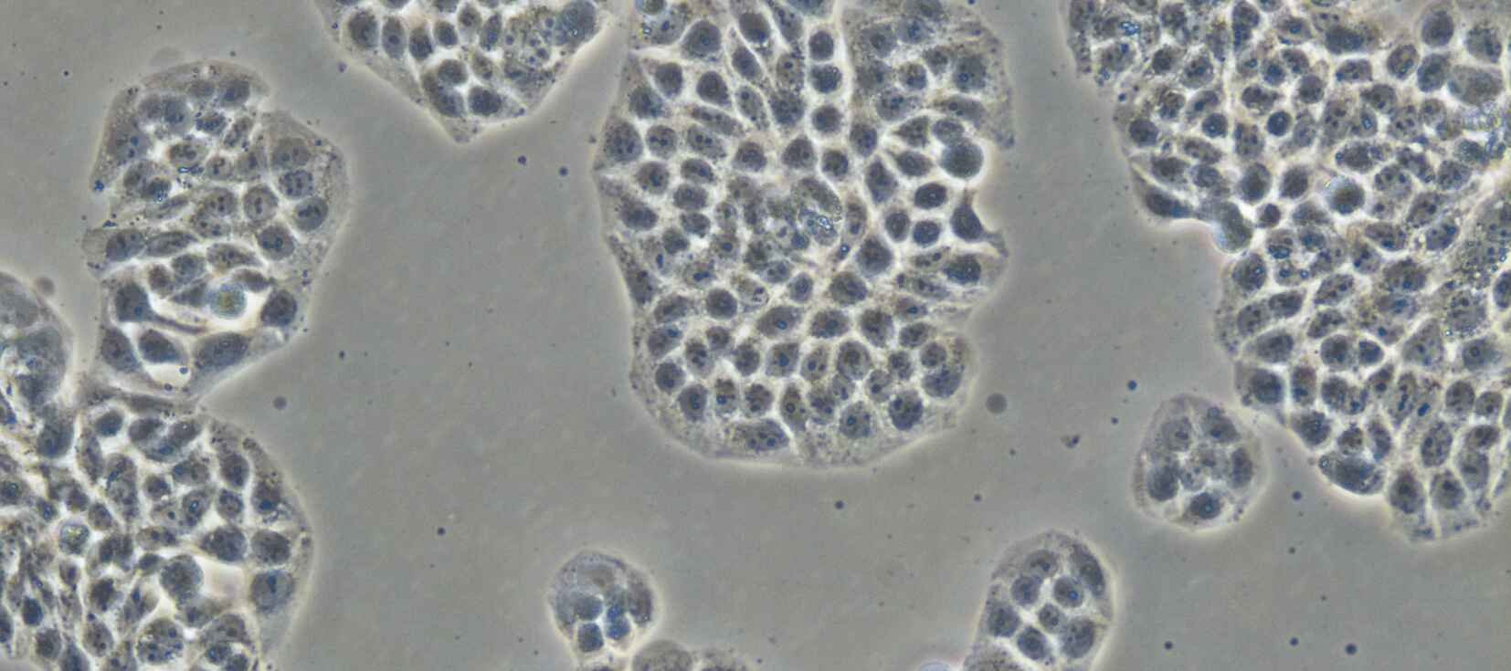
Standard Phase Contrast Image of adherent cells
Apodized Phase Contrast Image of the same group of cells
A wide variety of cell types are cultured in vitro, including primary cells recently obtained from tissue, immortalized cell lines, and stem cells. These different types of cells present similar imaging challenges.
An obstacle to seeing cells is their generally high degree of transparency, which restricts the usefulness of standard brightfield transmitted light microscopy. For this reason, tissue culture microscopes are often equipped for phase contrast imaging – a technique that uses interference to translate subtle differences in optical path length into contrast (even in mostly transparent specimens). Importantly, phase contrast is compatible with plastic culture vessels, unlike polarization-dependent techniques such as differential interference contrast (DIC).
Phase contrast makes identification of single cells relatively easy compared to brightfield, even at low magnification (objective lenses with a magnification between 4 – 20X are most often used for tissue culture). However, phase contrast imaging requires specialized objective lenses and a corresponding annular mask in the illumination light path. Furthermore, there are various types of phase contrast objectives to choose from.
Nikon’s CFI Achromat Series for Apodized Phase Contrast objective lenses are useful for tissue culture observation thanks to the provided reduction in the halo artifact that arises in areas with large optical path differences. This makes it easier to identify the borders of cells and to accurately observe larger model systems, such as tissues and whole organisms, compared to standard phase contrast objectives.
The optional Contrast Shield accessory blocks room light, providing an easy and cost-effective method for achieving high signal-to-noise fluorescence observation in a brightly lit culture room.
Fluorescence imaging is becoming an increasingly routine requirement in the tissue culture laboratory. For example, a common task is verifying the expression of fluorescent protein fusions by transfected cells – important for evaluating transfection efficiency, proper localization, and expression levels. Additionally, fluorescent stains are also widely used for visualizing key subcellular components (e.g. Hoechst dyes for staining nuclei in live cells).
The ECLIPSE Ts2-FL features up to three different user-selectable LED illumination modules which are built into the microscope. The microscope stores compatible LED modules and filter cube positions, and the last used brightness setting. Nikon also offers an optional Contrast Shield accessory to reduce background ambient light, a common problem in brightly-lit culture rooms.
Tissue culture work is often repetitive, resulting in a need for an ergonomically correct system that prioritizes ease of use and economy of motion. The ECLIPSE Ts2 controls are grouped in zones depending on if they are used for transmitted light illumination (left side of the microscope), epifluorescence (right), or both (center).
Tissue culture microscopes are often placed inside biosafety cabinets in order to promote a sterile environment and help protect cell health. Thus, a compact microscope can be advantageous for such work. Additionally, the stage is positioned low on the body, making repeated sample exchange more ergonomic to perform from a sitting position compared to microscopes with stages that are positioned higher up on the body.
If you’re interested in Nikon microscope products for automated stem cell culture assays, we invite you to visit our Cell Screening product page. We also invite you to learn more about Nikon microscope products for regenerative medicine.


