니콘인스트루먼트코리아 | Korea
- ko Change Region
- Global Site
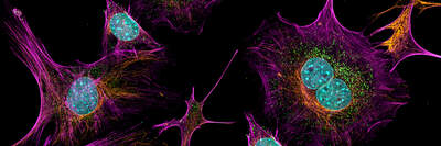
We provide imaging services for a variety of 2D model systems. These services include a broad portfolio of assays, live and fixed cell imaging on slide coverslips, dishes and multi-well plates, epifluorescence or confocal imaging modalities, and intelligent image acquisition and AI-supported analysis. Our broad portfolio of assays captures our strength, and are highly customizable to client-specific needs.
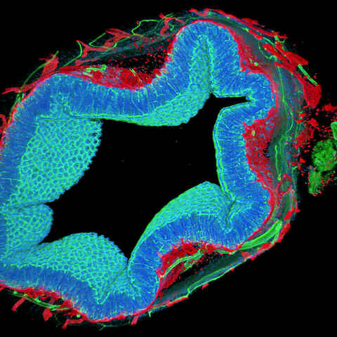
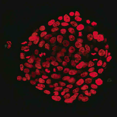
We provide imaging services for a variety of 3D model systems. These services include a broad portfolio of assays, live and fixed cell imaging, time-lapse imaging, whole organism imaging, epifluorescence or confocal imaging modalities, intelligent image acquisition, and AI-supported analysis. Our broad portfolio of assays captures our strength, and are highly customizable to client-specific needs.
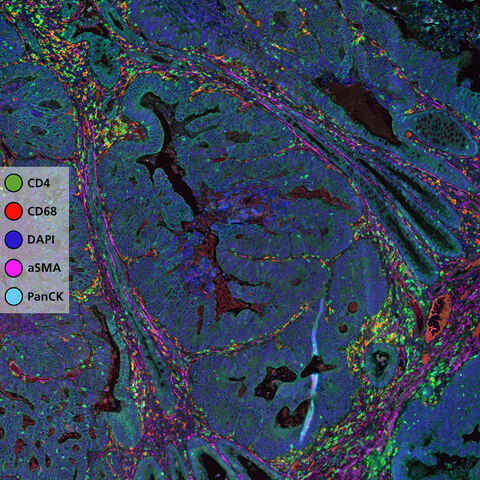
Advanced spatial biology technologies to perform high-plex spatial imaging and whole transcriptome spatial profiling. Assay functional markers, protein biomarkers, chemokines, cytokines and RNA within tissues, all while retaining spatial context.
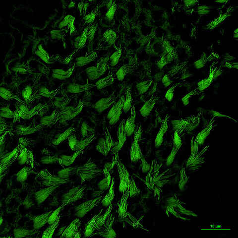
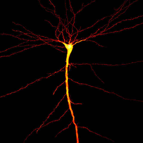
Unlock the power of ultra-detailed cellular imaging with super-resolution technology. Achieve unmatched clarity, ultra-low noise, and heightened sensitivity—ideal for revealing fine biological structures that standard microscopy can’t resolve.
Single molecule fluorescence in situ hybridization (smFISH), known for its ability to quantify individual RNA molecules, is widely used to characterize spatio-temporal patterns of gene expression in single cells. At NBIL we harness this technique to provide RNA quantification services for research applications such as gene expression analysis in response to drug candidates, spatial biology studies, and more.
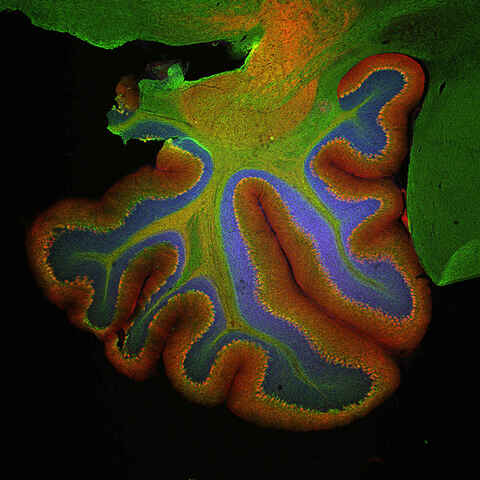
We provide whole slide imaging services, brightfield or fluorescence, of fixed specimens for histology, pathology, gene expression, and spatial biology studies.
Comprehensive imaging-based approaches for your toxicology studies during early drug development, drug safety assessments, and studies in MoA. Assess compound efficacy and efficiency in 2D and 3D cell culture models.
Customizable, automated, bias-free image acquisition and analysis for histology and pathology applications utilizing multiplexed chromogenic and fluorescent IHC, IF and ISH.
Generate consistent, high-quality data with Nikon’s turnkey automated microscopy and image analysis solutions.
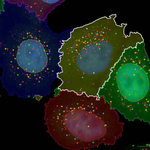
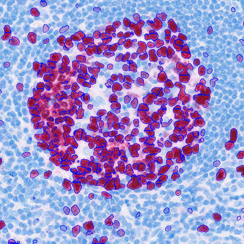
Image processing has a wide variety of uses and is an integral part of most microscopy experiments, from clarifying low-signal images to preparing images for robust segmentation and quantification. Custom analysis workflows are used for applications from gathering single-cell statistics to training AI networks for detection of customer-defined features.