니콘인스트루먼트코리아 | Korea
- ko Change Region
- Global Site
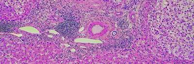
Customizable, automated, bias-free image acquisition and analysis for histology and pathology applications utilizing multiplexed chromogenic and fluorescent IHC, IF and ISH.
Tools for generating multiplex labeled chromogenic and fluorescent IHC, IF and ISH samples are abundantly available and used with increased frequency in the fields of histology and pathology. However, comprehensive and automated image acquisition and analysis solutions for R&D applications are not widely available to every researcher.
The Nikon BioImaging Lab offers customized, automated imaging and analysis services to help clients obtain robust, quantitative data from cell and tissue specimens that are labeled with chromogenic or fluorescent dyes and probes. The NBIL employs AI-assisted imaging analysis solutions to develop segmentation tools and workflows that can be customized to specific sample specifications.
