Nikon Europe B.V. | Europe & Africa
- fr Change Region
- Global Site
mars 2023
In drug development, two-dimensionally cultured monolayer cells have been used to evaluate the efficacy of a drug, measure effective concentration, and analyze mechanisms of action, through the use of cells. However, the use of two-dimensional (2D) cells for these purposes is problematic in that the cells show a different drug response or resistance to that of the living organism, so correct evaluation is difficult. On the other hand, cellular aggregates such as spheroids are known to exhibit reactions similar to those of the organism by forming the three-dimensional (3D) structure (reference 1). For this reason, the use of spheroids for pharmacological and toxin evaluation has increased in recent years. In addition, with spheroids, characteristics and physicochemical environments vary depending on the differences in shape and structure of the outer and inner layers (reference 2), so information on positional relationships in the three-dimensional structure is also important to analyze drug efficacy. Consequently, the acquisition of clear 3D images, high throughput for imaging a large number of samples, and analysis of 3D images are essential for effective and efficient analysis of spheroids using a microscope.
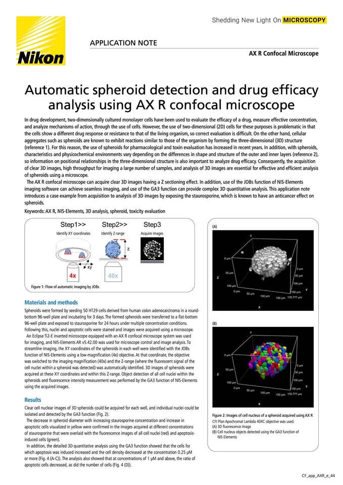
The AX R confocal microscope can acquire clear 3D images having a Z sectioning effect. In addition, use of the JOBs function of NIS-Elements imaging software can achieve seamless imaging, and use of the GA3 function can provide complex 3D quantitative analysis. This application note introduces a case example from acquisition to analysis of 3D images by exposing the staurosporine, which is known to have an anticancer effect on spheroids.