


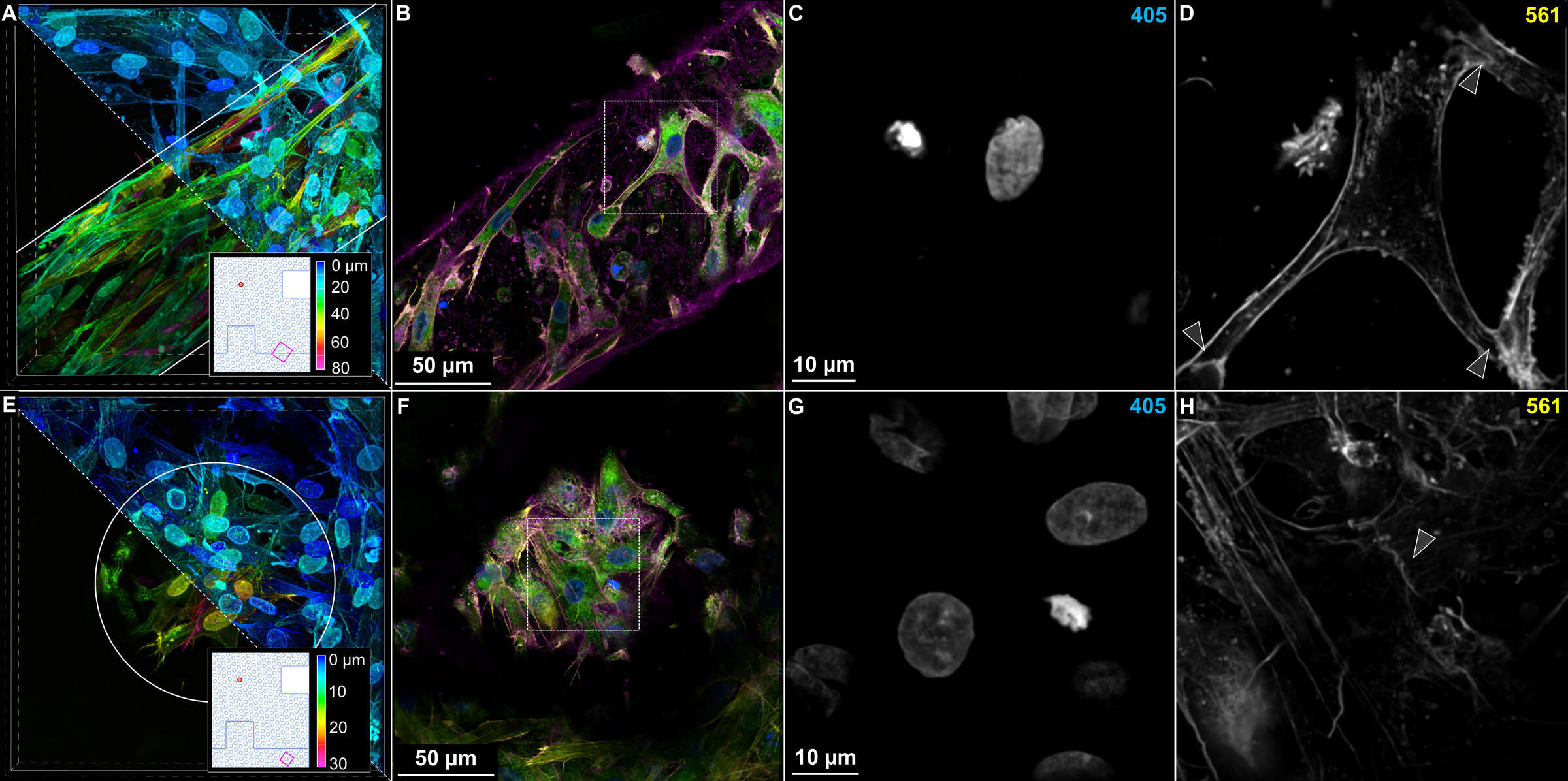
Figure 5: Cell-cell support behavior fostered by the Bio-Block microenvironment. (A) Depth-coded 3D view of cells proliferating at the block-block interface. The 221 x 221 x 100 μm volume was imaged in resonant scanning mode using the 40x Plan Apo λS silicone immersion objective and NSPARC detector. In total, 201, 1024 px frames were collected over 120 seconds. The cutaway reveals plane of interest 18 μm beneath the surface cell layer. Solid gray lines indicate underlying Bio-Block structure. (B) Plane of interest featuring a variety of cell-cell contacts commonly seen in Bio-Block cell culture. 4 μm EDF. (C, D) Isolated nuclear and cytoskeletal channel views from boxed region in B. Arrowheads indicate points of cell-cell contact. (E) Depth-coded 3D view of cells proliferating at the block-block interface. The 204 x 204 x 32 μm volume was imaged in galvo-scanning mode using the 40x Plan Apo λS silicone immersion objective and NSPARC detector. In total, 39, 1024 px frames were collected over 40 seconds. The cutaway reveals plane of interest 10 μm beneath the surface cell layer. Solid gray circle indicates underlying microchannel structure. (F) Plane of interest featuring a variety of cell-cell contacts commonly seen in Bio-Block cell culture. 4 μm EDF. (G, H) Isolated nuclear and cytoskeletal channel views from boxed region in E. Arrowheads indicate points of cell-cell contact.