


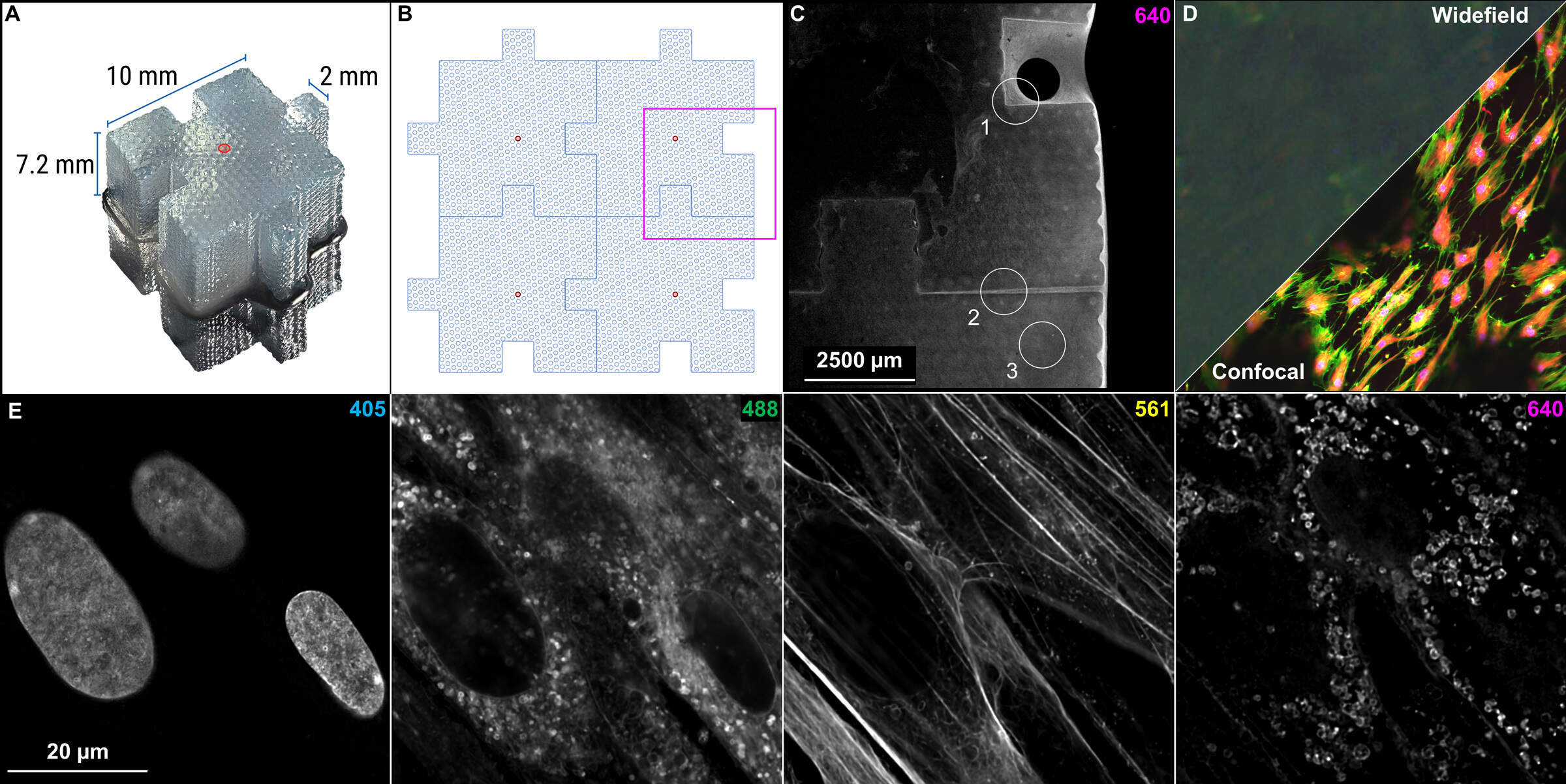
Figure 1: Bio-Blocks enable 3D, tissue-mimetic cell culture. (A) Macro image of a Ronawk Bio-Block with central microchannel highlighted in red. (B) Diagram of interconnected Bio-Blocks and microchannel locations. (C) Interconnected Bio-Blocks imaged with a 2x Plan Apo λD objective lens on the AX R and zoomed to the approximate boxed region in panel B. Three regions of interest are highlighted: 1, where cells have proliferated into a block intrusion; 2, where cells have proliferated within a block-block interface; and 3, where cells have started to proliferate into one of the printed microchannels. (D) Comparison of widefield epifluorescence imaging (top left of diagonal line) versus confocal imaging (bottom right of diagonal line). (E) Cells in this application note were labeled with Hoechst 33342 to stain nuclei (405), Memglow™ 488 to stain the lipid membranes (488), phalloidin Texas Red to stain filamentous actin (F-actin) in the cytoskeleton (561), and wheat germ agglutinin Alexa Fluor™ 647 to stain glycoproteins in the plasma membrane (640). Cells were cultured for 14 days before fixation. Image acquired with the 40x Plan Apo λS silicone immersion objective and NSPARC array detector.