Nikon Europe B.V. | Europe & Africa
Artboard 1
fr Change Region

Global Site

Princeton University

Contactez

Centre d’excellence

Princeton University

Princeton’s Nikon Center of Excellence was established to provide cutting-edge imaging technology to the local scientific community and to establish a mechanism for the exchange of ideas, methodologies and technologies between Princeton’s investigators and Nikon’s technology development teams. 

The Center enables users to investigate the dynamics of single molecules to whole organisms and tissues by providing state-of-the art confocal and super-resolution imaging systems and expert technical guidance and support from the Center’s Director.

Contactez

CofE Director

email hidden; JavaScript is required

Address

Princeton University
Department of Molecular Biology
116A Moffett Laboratory
Washington Road,
Princeton, NJ 08544
(609) 258-5432


Systems Available

N-STORM/TIRF/CSU-21 Super-Resolution & Spinning Disk Confocal System

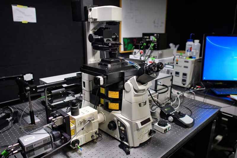
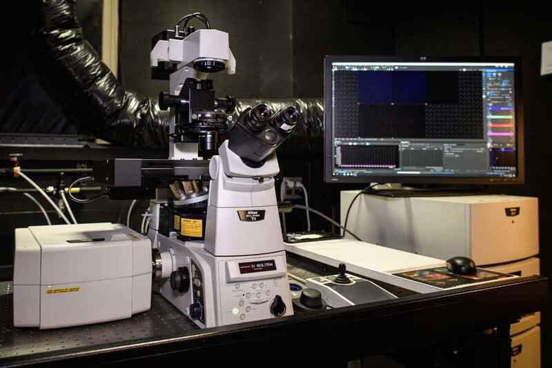
This system combines super-resolution N-STORM single molecule imaging with spinning disk confocal microscopy and ablation to provide a multimodal instrument suited for many biological imaging applications.

Components

  • Ti-E inverted microscope with Perfect Focus System (PFS)
  • N-STORM super-resolution single molecule localization microscopy system
  • 4-line laser unit (405nm, 488nm, 561nm, 647nm)
  • Andor iXon 897 EMCCD camera
  • Platine Z piezo série Mad City Labs Nano-Z
  • Système d'incubation par étapes Okolab
  • Andor Zyla 4.2 sCMOS camera
  • Rapp OptoElectronic UGA Firefly Ablation Module
  • Yokogawa CSU-21 spinning disk confocal
  • NIS-Elements software

CSU-21 Spinning Disk Confocal System

Fast multichannel confocal imaging is realized by combining spinning disk confocal scanner with emission splitter, with stimulation additionally provided using a galvo-based system.

Components

  • Ti-E inverted microscope with Perfect Focus System (PFS)
  • Yokogawa CSU-21 spinning disk confocal
  • Andor TuCam two-camera adapter for simultaneous two-color imaging
  • Hamamatsu ORCA-Flash4.0 V2+ sCMOS camera
  • Hamamatsu ImagEM EMCCD camera
  • Large format DS-Ri2 color CMOS camera
  • Platine Z piezo série Mad City Labs Nano-Z
  • Système d'incubation par étapes Okolab
  • 4-line laser unit (405nm, 488nm, 561nm, 640nm)
  • Bruker Mini-Scanner galvo-based stimulation system
  • Anton-paar 502 Rheometer
  • NIS-Elements software

Système à super-résolution N-SIM

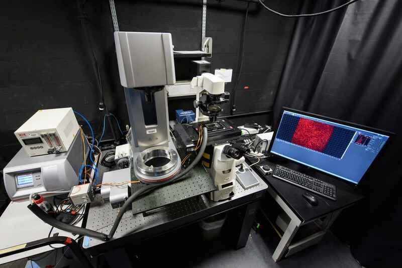
This N-SIM structured illumination super-resolution system beats the diffraction limit of resolution by a factor of two and features emission splitter for simultaneous two-color imaging.

Components

  • Ti-E inverted microscope with Perfect Focus System (PFS)
  • N-SIM super-resolution structured illumination microscopy system
  • Two-camera emission splitter for simultaneous 2-color imaging
  • 2x Andor iXon 897 EMCCD camera
  • Tokai Hit stage-top incubation system
  • NIS-Elements software

A1Rsi Resonant-Scanning Confocal System

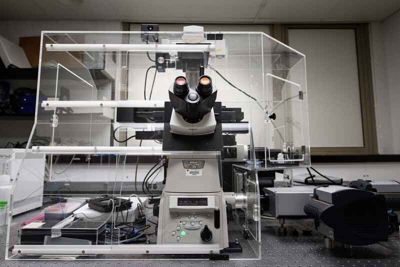
This A1R HD resonant scanning confocal system features both a spectral detector for linear unmixing of multiple sisgnals and DMD for patterned stimulation, perfect for applications such as optogenetics.

Components

  • Ti-E inverted microscope with Perfect Focus System (PFS)
  • A1R HD high-definition resonant scanning confocal system
  • A1-DUS 32-channel spectral detector
  • LUN-4 4-line laser unit (405nm, 488nm, 561nm, 640nm)
  • DU4G GaAsP 4-channel confocal detector
  • Platine Z piezo série Mad City Labs Nano-Z
  • Warner Instruments incubation system
  • Mightex Polygon400 DMD for patterned illumination (Lumencor AURA II light engine)
  • Andor Zyla 4.2 sCMOS camera
  • NIS-Elements software

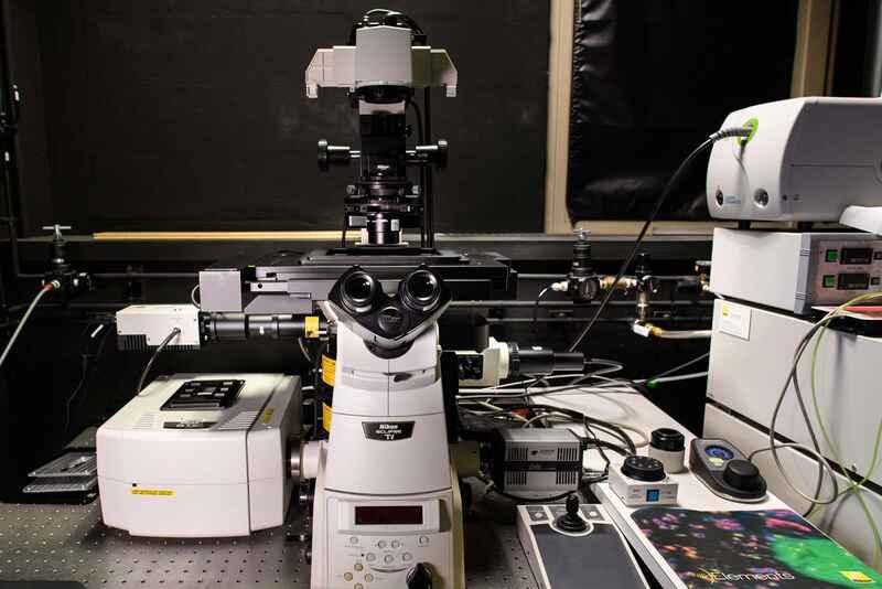
Go-to A1 point scanning confocal system configured on Ti-E inverted microscope, great for a wide variety of applications.

Components

  • Ti-E inverted microscope with Perfect Focus System (PFS)
  • 4-line laser unit (405nm, 488nm, 561nm, 633nm)
  • DU4G GaAsP 4-channel confocal detector
  • NIS-Elements software

Inverted Widefield Microscope System

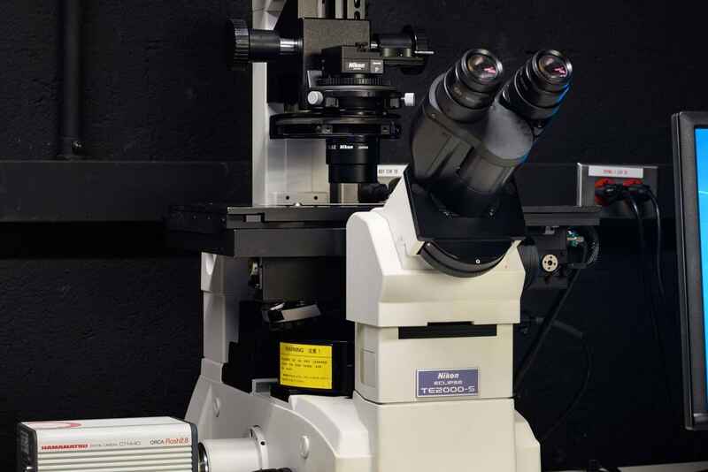
Inverted widefield system configured on TE2000 for routine applications.

Components

  • TE2000 inverted microscope
  • Hamamatsu ORCA-ER CCD camera