
Enduring Excellence - Long-Term Performance in Practice at VIB-KU Leuven
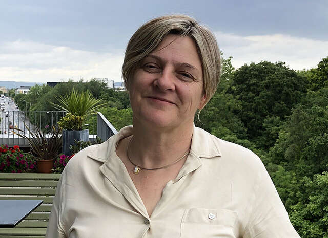
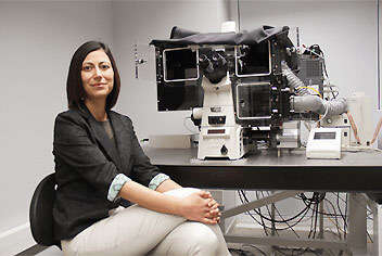
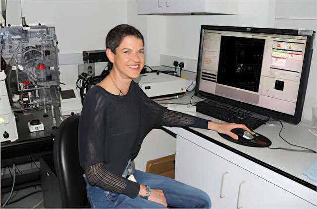
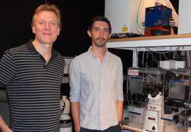
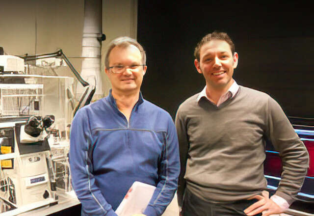
Nikky Corthout
Light Microscopy Expert - VIB BioImaging Core Leuven, VIB-KU Leuven Center for Brain and Disease
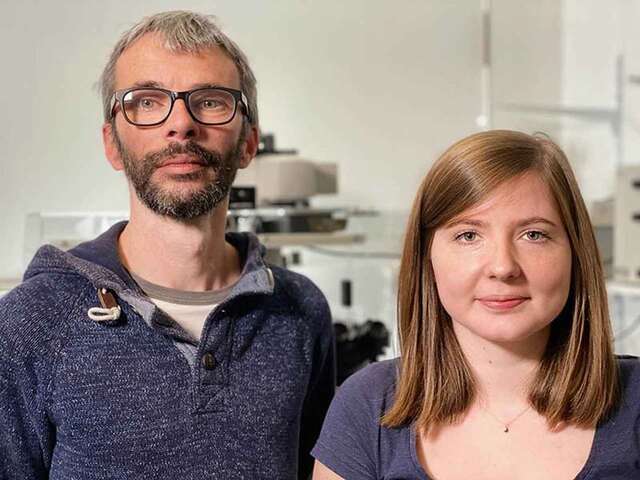
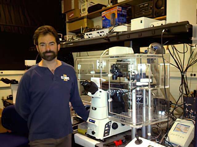
Pablo Hernández Varas
Head of Core - VIB BioImaging Core Leuven, VIB-KU Leuven Center for Brain and Disease
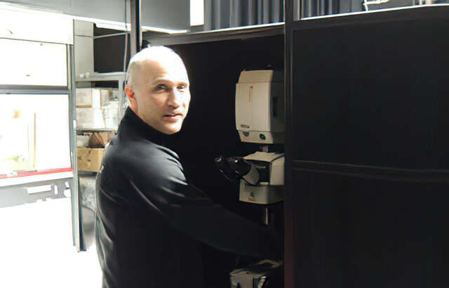
For more than a century, Nikon microscope systems have been built to last, and the stories behind their longevity continue to inspire us. In this edition of our Enduring Excellence series, we talk with Pablo Hernández Varas and Nikky Corthout, from the VIB BioImaging Core Leuven at Nikon Center of Excellence at KU Leuven, who have been using the Nikon A1 R confocal microscope system that’s been with the lab for over 17 years. Their insights highlight not just the system’s durability, but how it continues to support cutting‑edge scientific discovery to this day.

“Automating adjustments has reduced wrist and arm strain, making long practice sessions much more comfortable.”
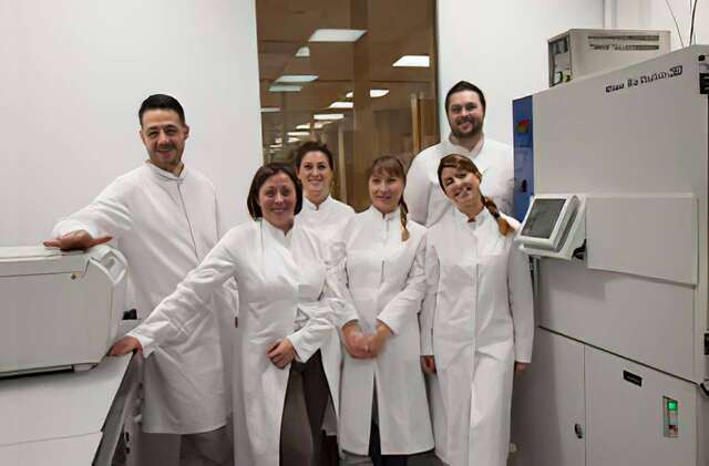
The ECLIPSE Ti2-I supports embryologists training at South Korea’s ART Academy
Eun-Kyung Kim, Ph.D
CHA University Medical Center
Global CHA Embryolab Academy
Fertility Training Center Director
Declining birth rates are a major global issue, and the need for training of embryologists — specialists who handle oocytes, sperm, and fertilized embryos — has become increasingly urgent to help address this problem. The CHA Biomedical Group in South Korea has established the Global CHA Embryolab Academy (GCEA), a specialized institution that systematically trains embryologists to support assisted reproductive technology (ART).
At GCEA, Nikon’s ECLIPSE Ti2-I motorized inverted microscope for ICSI and IMSI plays a vital role for embryologists. Dr. Eun-Kyung Kim, a highly experienced embryologist and director of the Fertility Training Center at the institute, shared insights on the microscope’s operation and the benefits of its implementation.
*This article contains interviews with healthcare professionals regarding our products. However, these interviews do not guarantee the efficacy, effectiveness, or performance of the products, nor do they constitute an official endorsement, recommendation, advice, or selection by the featured professionals.

Sample preparation utilizing the SMZ18 is the key to microscopic videography
YONE PRODUCTION CO., LTD.
Kanako Morioka (Director, Research Department)
Preparing high-quality samples is the first step towards capturing beautiful images. Making samples for microscopy is a detailed, sometimes hours-long process that requires patience, skill, and ingenuity. Yone Production, a company that produces life science videos, employs the SMZ18 stereo microscope for this purpose. Here, Kanako Morioka of the Research Department at Yone Production, who oversees sample preparation, shares her experience of using the SMZ18.

YOUR SWITCH TO THE FUTURE
Department of Biomedicine, University of Basel
Pascal Lorentz
Dr. Michael Abanto
Director of the Nikon Center of Excellence
Head of the Microscopy Core Facility
Research Outline: Immunology and infectious diseases, neurosciences, cancer biology, and tissue development and regeneration.

Dr. Kevin Richetin
Our Nikon Microscopy Ambassador Dr. Kevin Richetin is a neuroscience researcher studying Alzheimer’s disease and the development of tools for neurodegenerative diseases. In this interview video, Dr. Richetin discusses how they chose the Nikon microscope because its software NIS-Elements allows seamless integration of analysis and automation. He emphasizes that this unique capability is incredibly valuable for tracking particles with extremely high resolution over extended periods, a feature that sets Nikon apart.

Customer Interview - A step up in digital pathology using innovative digital microscopy.
Kitasato University Kitasato Institute Hospital
Director, Department of Pathology
Dr. Ichiro Maeda
Currently there are various treatment methods under development that could provide optimal medical care in response to the pathological attributes of each patient. Therefore, the role required of pathological diagnosis, which is indispensable for the planning of treatment policies and strategies, is becoming more important than ever before. However, the shortage of trained pathologists who assume this role is a common issue, not only in Japan, but also worldwide. To solve this problem, digital pathology has been attracting attention. In this article, we asked Dr. Ichiro Maeda, Director of the Department of Pathology, Kitasato University Kitasato Institute Hospital, for his impressions of using the digital image display optical microscope "ECLIPSE Ui".
*Dr. Maeda has provided feedback to Nikon on this microscope’s features and clinical usefulness based on his own personal experience.

YOUR SWITCH TO THE FUTURE
Cincinnati Children’s Hospital Medical Center
Matthew Kofron, Ph.D.
Professor, UC Department of Pediatrics
Alicia Ostmann
Cincinnati Cystic Fibrosis Theratyping Research Center
Kentaro Iwasawa, M.D.
Takebe Lab, Division of Gastroenterology
Research Outline: Child health improvement. Organoid creation. Drug efficacy for cystic fibrosis patients.

YOUR SWITCH TO THE FUTURE
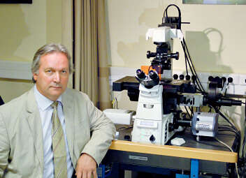
European Institute for Molecular Imaging (EIMI)
Professor Friedemann Kiefer, Ph.D.
Department of Intravital Molecular Imaging
Research Outline: Elucidation of how molecular mechanisms shape human cells and tissues biologically. Multiscale imaging.

YOUR SWITCH TO THE FUTURE
National Institutes of Natural Sciences, National Institute for Physiological Sciences
Tetsuhisa Otani, Ph.D. Assistant Professor
Division of Cell Structure
Research Outline: Epithelial tissue.

YOUR SWITCH TO THE FUTURE
Korea Advanced Institute of Science and Technology
Professor Won Do Heo, Ph.D.
Department of Biological Sciences
Research Outline: Optogenetic and bio-imaging technologies.

YOUR SWITCH TO THE FUTURE
Chinese Institute for Brain Research, Beijing, China
Dr. Hu Zhao, Principal Investigator
Research Outline: New tissue clearing methods, and building a neural connectome mapping platform based on these methods.

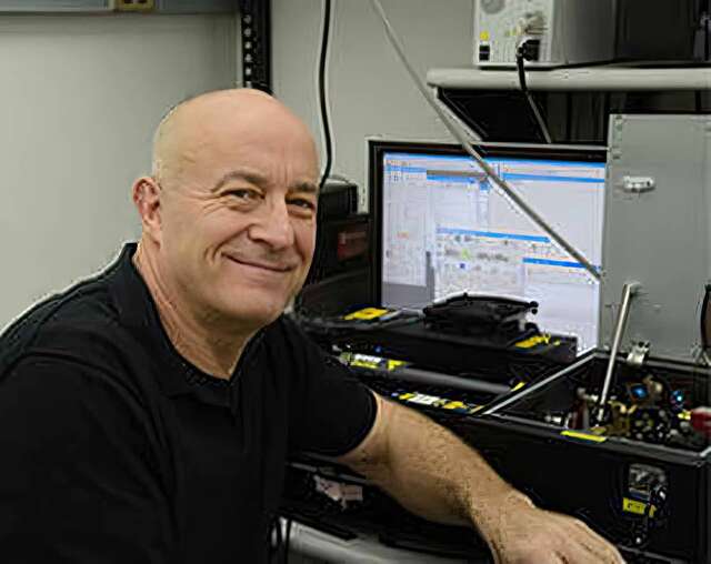
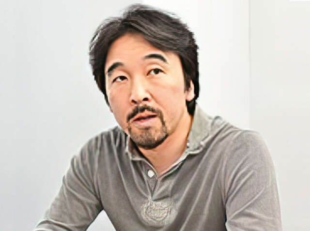
Customer Interview - “I believe this can change conventional imaging for pathologists”
Takeshi Sasaki
Professor, Project Leader, M.D. Ph.D
The University of Tokyo Hospital
Dept. of Next-Generation Pathology Information Networking
In medicine, pathologists play an extremely important role in confirming observation. They are often making observations with a microscope for long periods of time and the physical and mental burden of this is heavy. Aiming to reduce such stress issues for pathologists, Nikon has developed a new microscope for pathological examination, the ECLIPSE Ui, which allows pathologists to view high-resolution images of specimens on a monitor, rather than looking through eyepieces. Here, Dr. Takeshi Sasaki of the University of Tokyo Hospital, who has evaluated this microscope, talks about his impressions of it.
*Dr. Sasaki has provided feedback to Nikon on this microscope’s features and clinical usefulness based on his own personal experience.

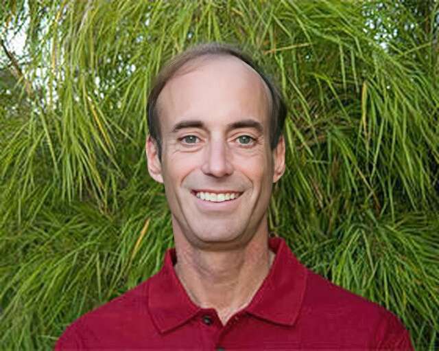
Nikon BioImaging Lab contributes to “mini-gut” research
Dr. Hidenori Akutsu
Director of the Department of Reproductive Medicine, Center for Regenerative Medicine, National Center for Child Health and Development
Nikon Equipment and Services

YOUR SWITCH TO THE FUTURE
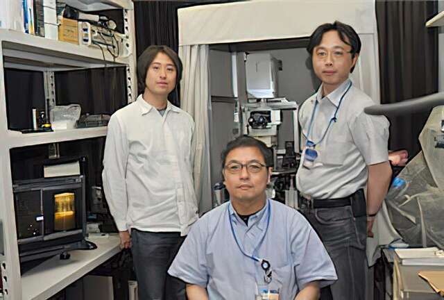
Research Institute for Microbial Diseases (RIMD) at Osaka University
Hiroaki Miki, Professor
Yosuke Funato, Associate Professor
Osamu Hashizume, Assistant Professor
Division of Cellular and Molecular Biology, Department of Cellular Regulation
Research Outline: Analyzing the function of a membrane protein molecule called Cyclin M, which plays a role in ejecting magnesium ions from cells.

Soutenir les études en classe avec l'ECLIPSE Ei
"Il est facile d'enseigner aux étudiants comment l'utiliser, et ils le trouvent également simple à utiliser"
L'observation à l'aide de microscopes joue un rôle extrêmement important dans l'apprentissage des bases de la science. L'ECLIPSE Ei de Nikon a été développé en tant que microscope à usage pédagogique avec le concept d'être un « microscope convivial même pour les étudiants qui l'utilisent pour la première fois ». Nous avons rendu visite à Mme Rika Izumi, professeur de sciences à l’école secondaire de premier et deuxième cycle de Rikkyo Niiza, Saitama, Japon, où l'ECLIPSE Ei est disponible dans les salles de classe, pour en savoir plus sur le contexte et les raisons du choix de ce microscope, l'expérience utilisateur et les réactions des élèves.

L'ECLIPSE Ci-L plus contribue à des examens cliniques plus confortables.
“Avec la haute performance optique, j'ai réalisé que sa conception réfléchie facilite notre travail quotidien“
Les examens cliniques jouent un rôle important dans les soins médicaux. Nikon a développé un nouveau microscope biologique, l'ECLIPSE Ci-L plus, avec le concept de réduire la fatigue physique et mentale des cliniciens et des techniciens de laboratoire qui utilisent quotidiennement des microscopes. Dans cette entrevue, nous avons parlé au Dr Akira Yoshikawa du Département de pathologie anatomique du Kameda Medical Center - un hôpital phare dans la partie sud de la préfecture de Chiba, au Japon - de ses réflexions après avoir utilisé ce microscope et l'objectif nouvellement développé pour les microscopes 'CFI Plan Apochromat Lambda D' pour une utilisation pratique au quotidien.
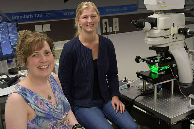
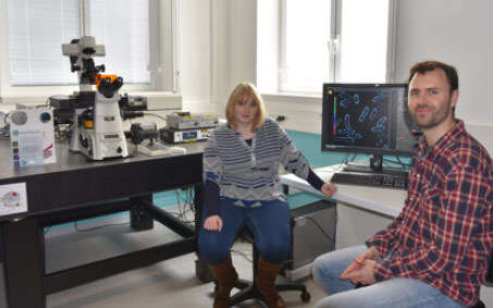
Associate Prof. Dr. Klazina Kooiman and Dr. Ines Beekers
Associate Prof. Dr. Klazina Kooiman, Head of Therapeutic Ultrasound Contrast Agent Group, and Dr. Ines Beekers, Postdoctoral Researcher in the Department of Biomedical Engineering of the Thoraxcenter, Erasmus MC, Rotterdam, the Netherlands
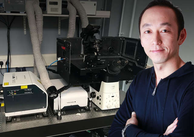
Dr. Yohei Yamauchi
Dr. Yohei Yamauchi, Principal Investigator, Cell biologist of viral infections, School of Cellular and Molecular Medicine, University of Bristol, UK
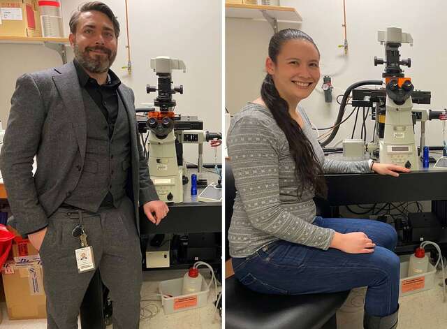
Assistant Prof. Joseph Michael Hyser & Dr. Alexandra Leigh Chang-Graham
Virology and Microbiology
Baylor College of Medicine
Houston, Texas, USA
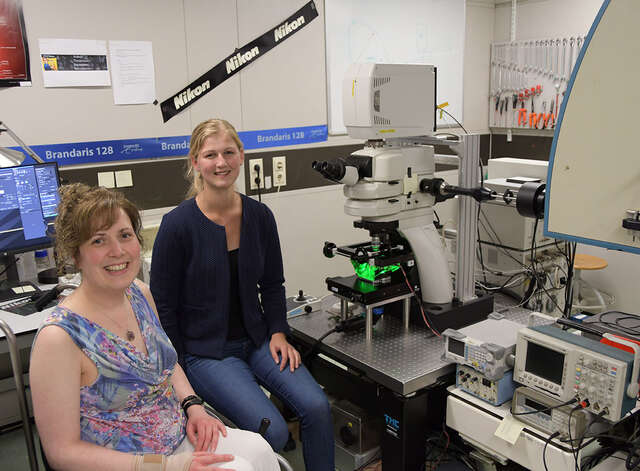
Assistant Prof. Klazina Kooiman & Inés Beekers
Therapeutic Ultrasound Contrast Agent Group, Thoraxcenter, Department of Biomedical Engineering, Erasmus MC, Rotterdam
Dr. Steven Nedellec and Dr. Tiphaine Douanne
Dr. Steven Nedellec, Facility Manager of MicroPICell, Université de Nantes, France and Dr. Tiphaine Douanne, Universite de Nantes, Signaling in Oncogenesis, Angiogenesis and Permeability, CRCINA INSERM U1232, France
Dr. Tadahiro Iimura
Division of Bio-Imaging, Proteo-Science Center (PROS), Ehime University
Division of Analytical Bio-Medicine, Advanced Research Support Center (ADRES), Ehime University
Graduate School of Medicine, Ehime University
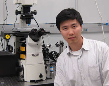
Melike Lakadamyali, Ph.D.
The advanced Fluorescence Imaging and Biophysics Group, ICFO-Institute of Photonic Sciences
Simon C. Watkins, Ph.D.
Professor and Vice Chairman of the Dept. of Cell Biology
Director and Founder of the Center for Biologic Imaging
University of Pittsburgh
Pittsburgh, Pennsylvania, USA
Ronald D. Vale, Ph.D.
Professor and Vice-Chairman of the Department of Cellular and Molecular Pharmacology
Investigator, Howard Hughes Medical Institute (HHMI)
The University of California, San Francisco San Francisco, CA, USA
Tomomi Nemoto, Ph.D. and Ryosuke Kawakami, Ph.D.
Research Institute for Electronic Science
Sapporo, Hokkaido, Japan
Tamas Freund, Ph.D. and Istvan Katona, Ph.D.
Institute of Experimental Medicine of the Hungarian Academy of Sciences (IEM HAS)
Budapest, Hungary
Maddy Parsons, Ph.D.
Group Leader (Royal Society University Research Fellow)
Randall Division of Cell and Molecular Biophysics
King’s College London
London, United Kingdom
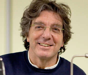
Alberto Diaspro, Ph.D.
Professor of Applied Physics, Department of Physics, University of Genoa
Director of the Department of Nanophysics, Istituto Italiano di Tecnologia
Paul Ronald Selvin, Ph.D.
Professor, Department of Physics, University of Illinois at Urbana-Champaign
Bo Huang, Ph.D.
Assistant Professor, Department of Pharmaceutical Chemistry, Department of Biochemistry and Biophysics, University of California, San Francisco
Atsushi Miyawaki, M.D., Ph.D.
Senior Team Leader, Laboratory for Cell Function Dynamics, RIKEN Brain Science Institute
Ikuo Wada, Ph.D.
Department of Cell Science, Institute of Biomedical Science, Fukushima Medical University
Romain Le Bars, Ph.D.
The Imagerie-Gif light microscopy core facility is a member of the France Bioimaging Infrastructure. The facility is hosted by the Institute for Integrative Biology of the Cell (I2BC) at Gif sur Yvette, France.
Prof. Staffan Strömblad, Ph.D.
Staffan Strömblad Ph.D. is group leader at the prestigious Karolinska Institutet, Stockholm, Sweden, an institution that awards the Physiology Nobel Prize yearly. He is also the head of the Live Cell Imaging Facility (LCI), in which the Nikon Center of Excellence for live cell imaging is integrate.
Dr. Arne Seitz, Dr. Romain Guiet and Thierry Laroche
The Faculty of Life Science (SV) at the École Polytechnique Fédéral de Laussane (EPFL), Switzerland, has a long record of excellence in research applied to life sciences
Masato Nakagawa
So-called iPS cells are attracting considerable interest as pluripotent stem cells that may open up a whole new world of medicine. The Center for iPS Cell Research and Application (CiRA) at Kyoto University is pursuing a wide range of research activities that aim to realize regenerative medicine utilizing iPS cells. The Nikon BioStation CT cell culture observation system is being used in this iPS cell research and is contributing to its efficiency.
We were pleased to have had an opportunity to speak with Masato Nakagawa, who is engaged in iPS cell research at CiRA.
Prof. Heinz Beck, Ph.D.
Full time professor at the Laboratory of Experimental Epileptology and Cognition Research hosted in the Life & Brain Center, Part of the University of Bonn.
Note: The institutions and job titles listed with each researcher reflect their affiliation at the time of the interview.
- Accueil
- Ressources
- Entretiens avec les clients