Nikon Instruments Inc. | Americas
- fr Change Region
- Global Site
Takeshi Sasaki
Professor, Project Leader, M.D. Ph.D
The University of Tokyo Hospital
Dept. of Next-Generation Pathology Information Networking
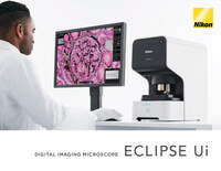
In medicine, pathologists play an extremely important role in confirming observation. They are often making observations with a microscope for long periods of time and the physical and mental burden of this is heavy. Aiming to reduce such stress issues for pathologists, Nikon has developed a new microscope for pathological examination, the ECLIPSE Ui, which allows pathologists to view high-resolution images of specimens on a monitor, rather than looking through eyepieces. Here, Dr. Takeshi Sasaki of the University of Tokyo Hospital, who has evaluated this microscope, talks about his impressions of it.
*Dr. Sasaki has provided feedback to Nikon on this microscope’s features and clinical usefulness based on his own personal experience.
Dr. Sasaki: At the University of Tokyo Hospital, there is a double-check system for observations, and we, as board-certified pathologists, double-check all the pathological observations that have been made by younger doctors. Then, we return the results if corrections are needed. It’s my turn to handle this task every Friday, and I’m responsible for checking all the samples delivered that day. I also utilize microscopes on other days in my lectures to medical students and when providing guidance for young teachers, but I really concentrate on using them on Fridays.
Dr. Sasaki: It depends on the number of samples received on that day, but I usually spend about eight to nine hours looking through microscopes throughout the day. In particular, the mammary gland is my specialty field, so I double-check all mammary gland samples taken at the University of Tokyo Hospital. When we have a lot of samples on Friday, it can often exceed nine hours. Besides, I am also the director of the pathology department, and support remote pathological observation at hospitals without pathologists, or that have only one pathologist, etc. So, all of this other work is added to the regular in-hospital observation time.
Dr. Sasaki: Since we pathologists look into microscopes while bending the body forward for extended periods of time, it puts a major burden on the body. And it’s not relaxing mentally. Usually, I don’t notice during observation because I’m fully concentrating, however, I often feel that it’s painful afterwards. My back hurts, and the joints of my body are stiff. In general, pathologists spend a considerable amount of time looking through microscopes during their daily work, so I guess other pathologists experience the same kinds of problems. We don’t get to walk around much either, so I certainly think it’s at the level of hard work.
Dr. Sasaki: When I used the ECLIPSE Ui, the most important benefit I felt was that it displays microscopic images of samples on the monitor in real time for observation. As a result, I didn’t have to hunker down to look into the eyepiece, which made everything much easier. In addition, since my face was raised, I experienced a sense of openness. Even if I continued with the observation for a long time, I didn’t feel constricted, and my impression was that the mental burden was considerably reduced
Dr. Sasaki: That the images of samples are digitalized is also a very important point. Until now, in pathological observation, when conducting conferences or consulting, we first read the samples with a scanner and create a digital image. However, with the ECLIPSE Ui, you can eliminate those steps. Just by setting the slide glass you can immediately acquire it as a digital image.
The ECLIPSE Ui and high-resolution monitor.
*The image on the monitor is a sample prepared by Nikon.
Observation of samples can be performed with a comfortably relaxed posture.
*The image on the monitor is a sample prepared by Nikon.
Dr. Sasaki: The startup after turning on the power and the response of the stage movement are very good. Also, the image display is quick, and there is no delay before the image is displayed on the monitor. It was all very smooth. The GUI is easy to understand, and I didn’t feel any stress.
Dr. Sasaki: I tried it with various samples, but this was developed by Nikon and the image is very clear. For example, it is difficult to confirm Helicobacter pylori with a digital image usually, but with this ECLIPSE Ui, the image was reproduced with the same accuracy as normal optical microscopes, without any stress. Also, since the macro image is displayed on the monitor, I can see exactly where I am currently observing at a glance, which is very convenient. There are colors that are easy to check depending on the samples. It was great that the colors could be easily adjusted via the GUI.
A macro image is displayed at the bottom right of the high-resolution monitor.
*The image on the monitor is a sample prepared by Nikon.
You can locate the area you are currently observing on the macro image at a glance.
*The image on the monitor is a sample prepared by Nikon.
Dr. Sasaki: The potential to change the image of pathologists, for a start. The conventional image was a somewhat gloomy impression of looking into microscopes with my back hunched over. However, now I can sit up and look in a more relaxed way at a monitor while making observations, for example, so I think trainee doctors and students who come to observe can gain a brighter impression. Also, this microscope can share digital images, and enables remote operation.
Dr. Sasaki: This microscope delivers a great advantage to my generation of pathologists, but it is even more useful for upcoming generations. Currently, practical training in pathology and histology in medical schools is mostly done using digital images. Trainees look at a monitor instead of peering through a microscope eyepiece. In the future, when the generation of so-called digital natives becomes the mainstream of pathologists, I think it will become common for multiple pathologists in remote locations to consult and make observations utilizing microscopes like the ECLIPSE Ui.
Dr. Sasaki: Today, digitization is essential in many areas of society. I strongly feel that the time has come for pathological observation to be digitized through next-generation microscopes such as the ECLIPSE Ui.
Note: The institutions and job titles listed with each researcher reflect their affiliation at the time of the interview.