Nikon Corporation Healthcare Business Unit | Asia, Oceania & Middle East
- en Change Region
- Global Site
Minimal experimental setup and complexity by AI-driven assays and analysis.
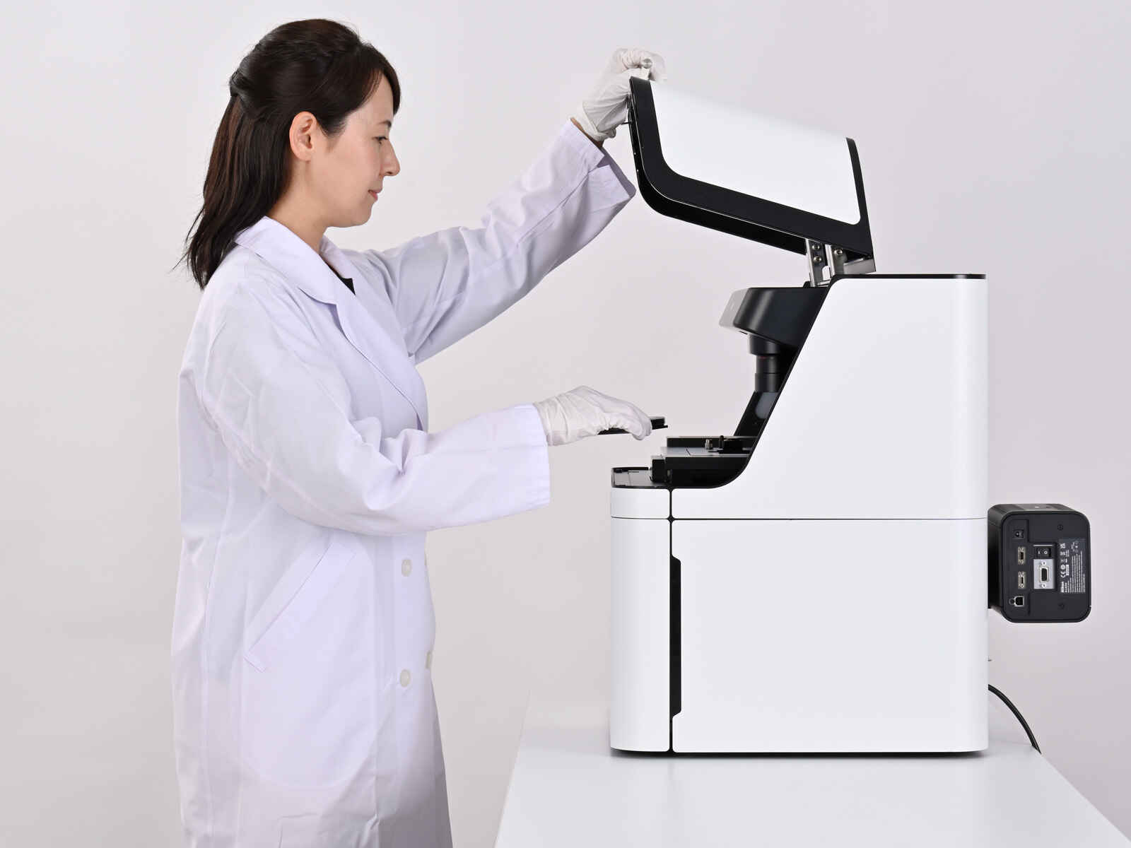
No need to master complicated microscope hardware and software – ECLIPSE Ji makes data collection and cellular imaging assays easy.
Renowned Nikon optics provide clear and sharp images on plate assay devices.
Utilizing Nikon’s precision optical hardware, all of the advantages of high sensitivity and resolution from a research-level microscope are retained by an AI-driven, easy-to-operate benchtop laboratory instrument.
Preconfigured and optimized turnkey assay experiments minimize time spent defining parameters, maximizing data collection.
Compares protein expression level changes in cells and cell nuclei in multiple wells.
Counts the number of cell nuclei in a fixed sample and the area of the well occupied by cells.
Measure changes in confluency and count nuclei in live samples over time.
Investigates the percentage of cells expressing the target protein, and reports the expression efficiency.
Analyzes cellular morphology with measurements of the cell nucleus, cytoplasm, and the size of the cell region.
Measures the percentage of dead cells among all cells and evaluates cytotoxicity.
Measures the nuclear translocation of NF-κB, indicating an extra-cellular stimulus.
Measures the number of autophagosomes, their area, and their fluorescence intensity.
Measures the number, area, and fluorescence intensity of bioparticles taken into the cell by phagocytosis.
Measures the number, area, and fluorescence intensity of granules formed by endocytosis, which are taken up from outside the cell.
Measures the number of cells containing micronuclei or multiple nuclei.
Measures the number, area, and fluorescence intensity of mitochondria.
Measure the number and length of neurites protruding from neuron cell bodies.
Measure the recovery of filled area in an artificially-created wound over time.
Measure the proportion of cells in each cell cycle phase based on Fucci, the genetically-encoded, fluorescent cell cycle reporter system.
ECLIPSE Ji’s Smart Experiment software interface uses newly developed artificial intelligence (AI), implemented to minimize errors and maximize data collection.
| Input | Output | ||
|---|---|---|---|
Load a plate | 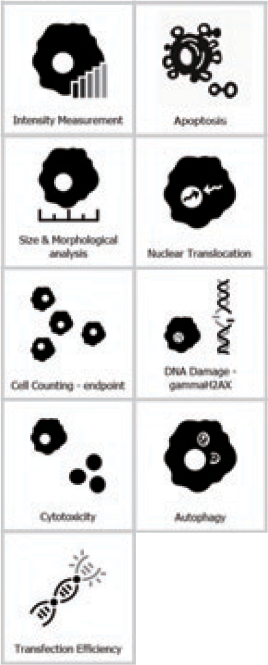
Select assay / Input basic information | 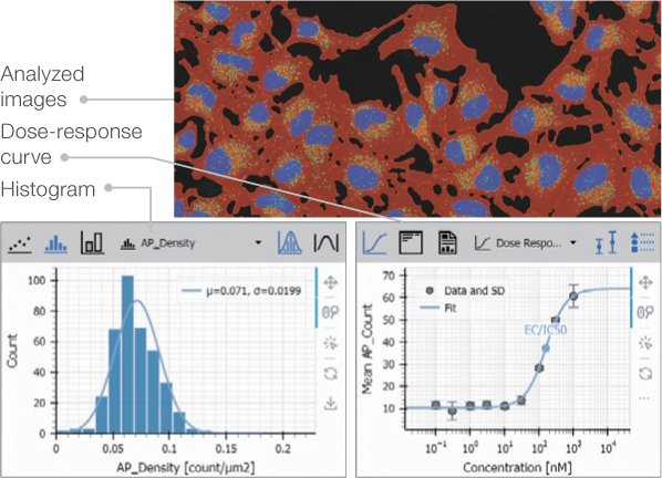
Check the shooting results and analysis results | 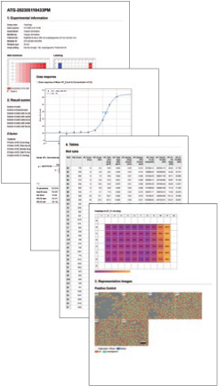
Report |
| step 1 | step 2 | step 3 | step 4 |
| ECLIPSE Ji | Pre-AcquisitionAI automatically judges plate status and automatically adjusts acquisition conditions |
Actual Acquisition/analysis/data displayAutomatic acquisition, data extraction, graphing under optimized acquisition conditions |
ReportingOne click export of the data |
AI based on Deep Learning defines acquisition settings and image analysis parameters, saving researchers valuable time at the microscope.
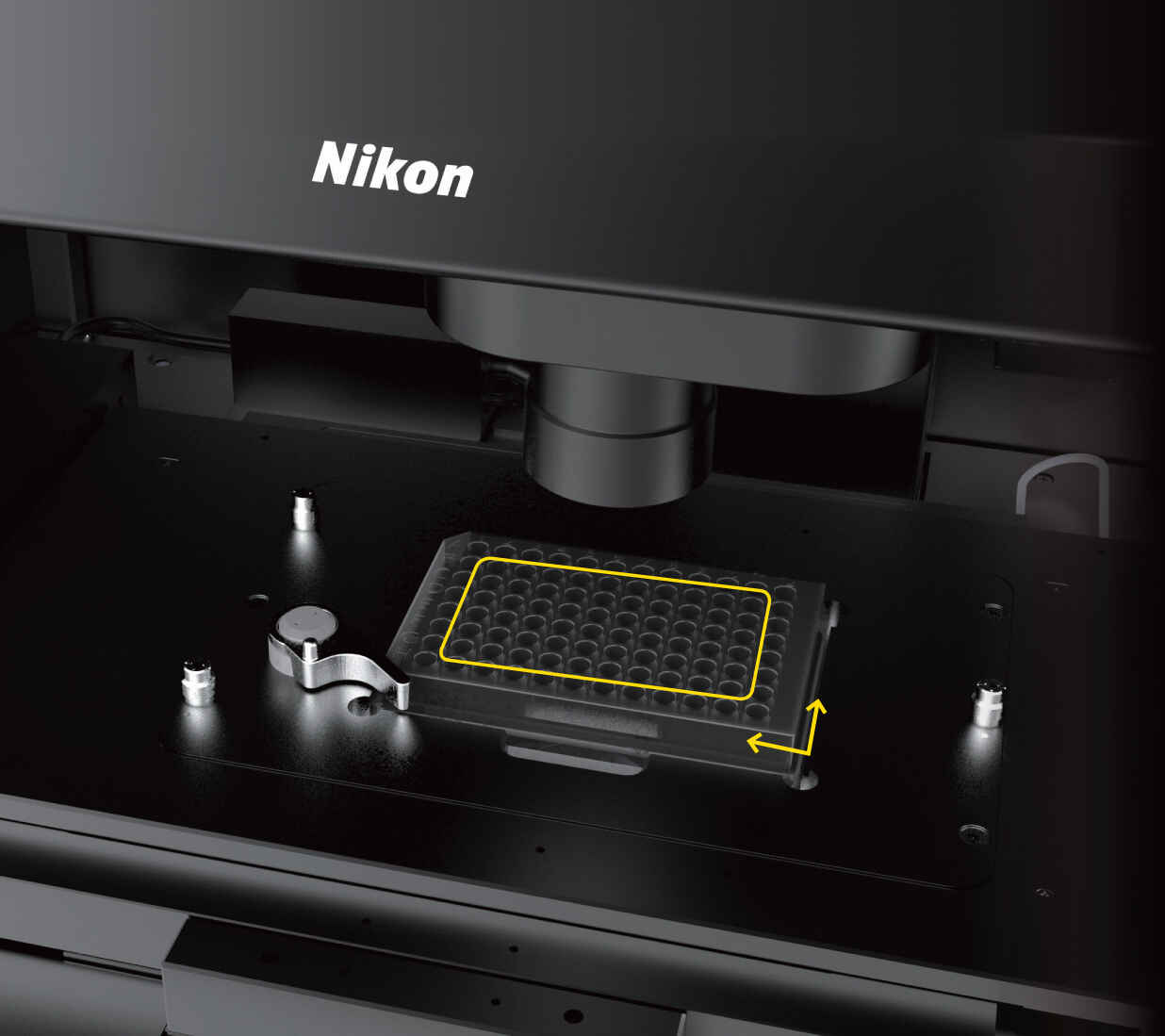
Plate type and dimensions are automatically detected. There is no need to select from lists or manually enter plate data.
A rapid preview across the entire plate determines which wells have sample present, allowing users to easily skip empty wells.
There is no need for troublesome tuning of light intensity and exposure time. The optimal exposure settings for image analysis are automatically calculated from the luminance values of all wells.
No alignment work is required. The system automatically detects and corrects the plate position.
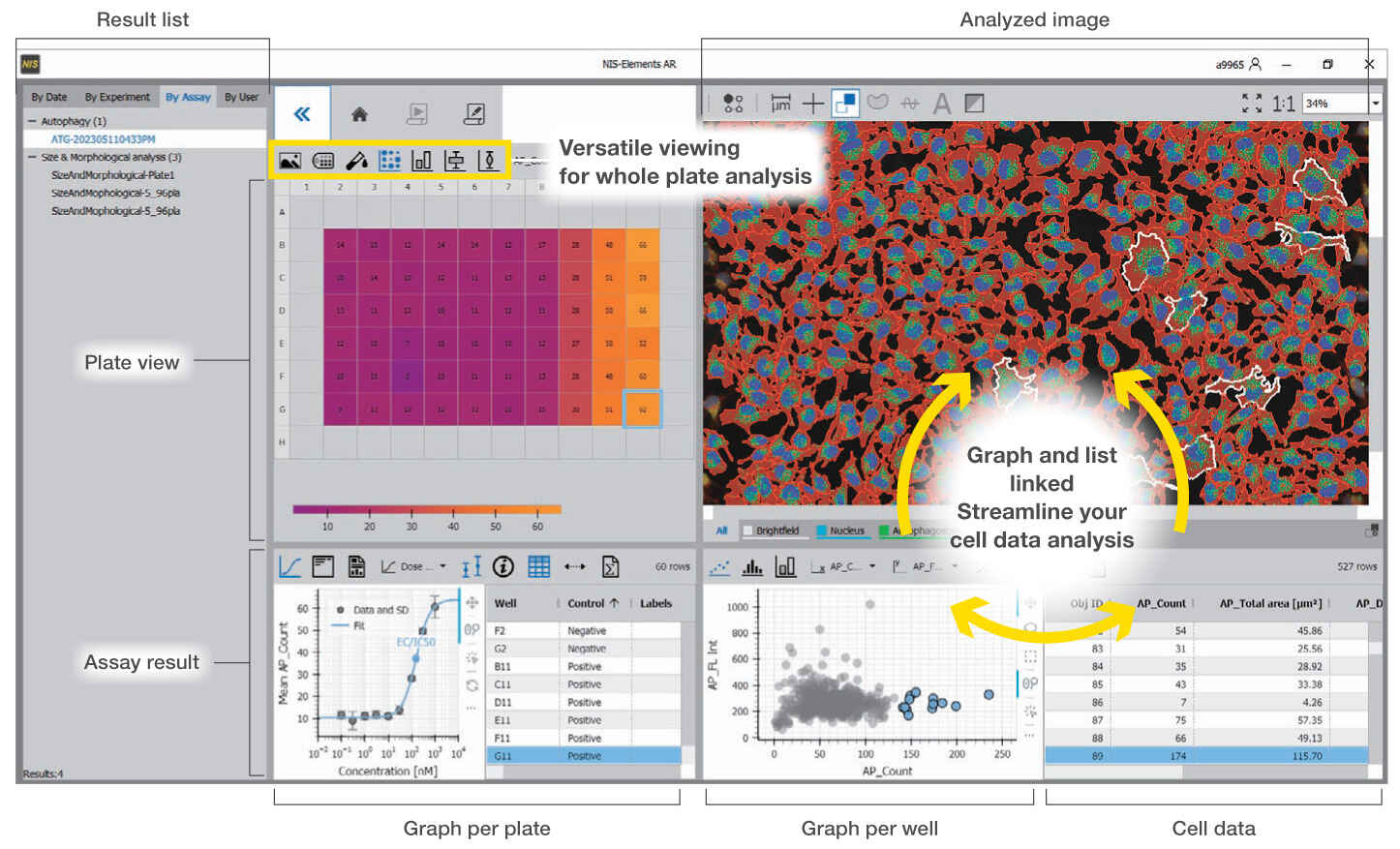
Images and corresponding analysis data for the plate, well, and each cell is contained in an interactive and linked interface. Users can navigate and quickly visualize trends and results.
Outside of plate assays, ECLIPSE Ji can also serve as a digital research microscope, and can be integrated with a variety of peripheral hardware ranging from filter wheels through confocal systems such as "AX", or high-sensitivity cameras.
*The design and specifications may differ from the actual product.