Nikon Europe B.V. | Europe & Africa
- de Change Region
- Global Site
September 2023
ECLIPSE Ji with Smart Experiment software enables seamless experiments from image acquisition to analysis and graph creation. Pre-trained Artificial Intelligence (AI) and pre-defined imaging processes automatically optimize image acquisition and analysis settings, providing visualized data and EC50 information with simple operation. Cytotoxicity analysis is a versatile assay used in a variety of biological research, such as studying the effects of pharmacological compounds, development of culture media and scaffolds for cell culture, or elucidation of cellular physiology. This application note introduces an example of using the Cytotoxicity module of Smart Experiment to visualize the dose-dependent induction of cell death due to staurosporine and quantify the effect of the drug by calculating the EC50.
Keywords: Cytotoxicity, Live / Dead, Cell viability, automatic setting, EC50, dose-response curve
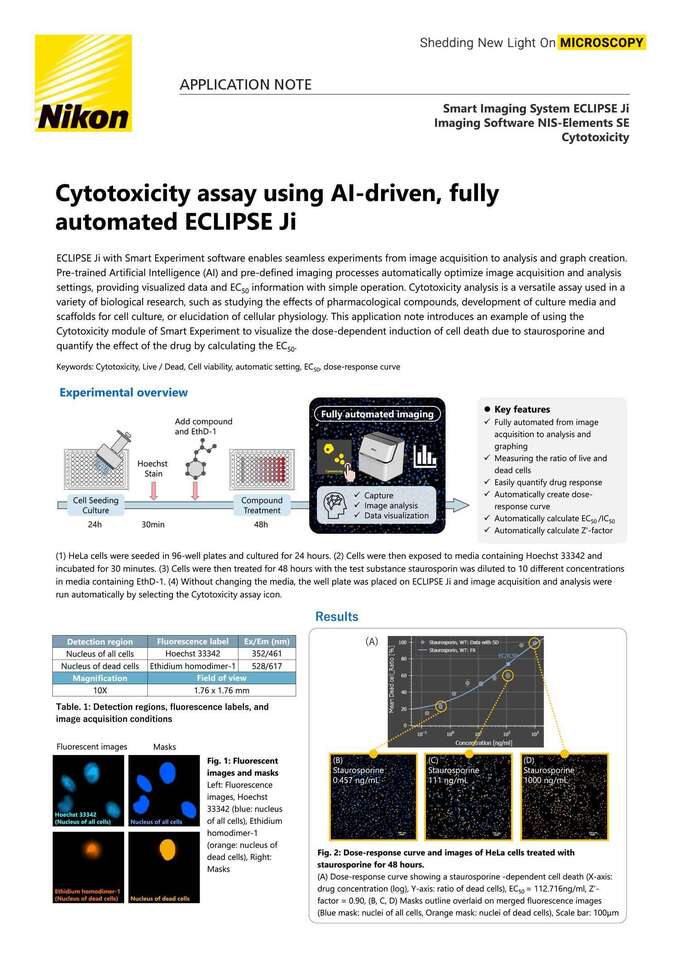
(1) HeLa cells were seeded in 96-well plates and cultured for 24 hours. (2) Cells were then exposed to media containing Hoechst 33342 and incubated for 30 minutes. (3) Cells were then treated for 48 hours with the test substance staurosporin was diluted to 10 different concentrations in media containing EthD-1. (4) Without changing the media, the well plate was placed on ECLIPSE Ji and image acquisition and analysis were run automatically by selecting the Cytotoxicity assay icon.
Fig. 2: Dose-response curve and images of HeLa cells treated with staurosporine for 48 hours.
(A) Dose-response curve showing a staurosporine -dependent cell death (X-axis: drug concentration (log), Y-axis: ratio of dead cells), EC50 = 112.716ng/ml, Z’-factor = 0.90, (B, C, D) Masks outline overlaid on merged fluorescence images(Blue mask: nuclei of all cells, Orange mask: nuclei of dead cells), Scale bar: 100μm
Fig.3: Analysis results
(A)Heat map showing ratio of dead cells
(B) Bar graph showing the number of cells for each well (blue: number of total cells, orange: number of dead cells, green: number of live cells), The plate map view display allows intuitive confirmation of the drug reaction in each well.
HeLa cells are seeded in a 96-well plate at a density of 6x10e3 cells/well and culture for 24 hours at 37℃ in a 5% CO2 incubator. Cells were stained with media containing 2 μg/ml Hoechst 33342 for 30 minutes at 37℃ in a 5% CO2 incubator. Staurosporin diluted in FluoroBright DMEM contained 4μM EthD-1 at concentrations of 0 nM, 0.152 nM, 0.457 nM, 1.41 nM, 4.11 nM, 12.3 nM, 37 nM, 111 nM, 333 nM, 1000 nMwas added to 6 replicate wells. The cells are treated staurosporin for 48 hours at 37℃ in a 5% CO2 incubator. To prevent detachment of dead cells, well plates were placed on ECLIPSE Ji without changing media to run image acquisition and analysis.
Cytotoxicity assay is an assay that measures the ratio of live and dead cells.Generally, DAPI cannot stain the nuclei of living cells. Please stain the nuclei of all cells with Hoechst 33342, not DAPI. In addition, washing after drug treatment detaches dead cells from the bottom of the well plate, reducing the accuracy of quantitative analysis. Therefore, image acquisition should be performed without washing or fixing the cells after drug treatment.
Compatible vessel*
・ 24, 48, 96 well plate
*Compatible with glass and polystyrene bottom well plates.If cell adhesion to the bottom is a priority over image quality, use well plates with polystyrene bottoms.
Chae, HJ, et al., Molecular mechanism of staurosporine-induced apoptosis in osteoblasts. Pharmacol Res 42 (4):373-81 (2000).
ECLIPSE Ji is an AI-Driven, fully automated imaging system. By using it in combination with NIS-Elements SE, image acquisition, analysis, and graph creation can be run seamlessly and automatically. It is equipped with "CellFinder.ai“, which uses AI to find the optimal focal plane in autofocus settings that normally require advanced human judgment. Various trained AIs are implemented in the image acquisition and analysis process. This greatly reduces the number of steps for setting and optimization and makes it easier for everyone to get results.
Imaging Software NIS-Elements SE
SmartExperiment Basic Set
Cytotoxicity


