![](https://downloads.microscope.healthcare.nikon.com/production/imager/mastheads/2210/LV100ND-POL-DS-Masthead_f076438097b2d2d85c6c3ef04742411e.jpg)
Mikroskope zur Dispersionsfärbung ermöglichen eine qualitative Analyse und Identifizierung von Asbest
Polarisierte Lichtmikroskope können die Eigenschaften von Asbest wie Brechungsindizes, Doppelbrechung, Verzögerung, Extinktionswinkel, Pleochroismus und Dehnungszeichenmessen, was bei der Identifizierung von Asbest hilft. LV100ND POL / DS ist ein Hochleistungs-Polarisationslichtmikroskop mit Zubehör, das eine Beobachtung der Dispersionsfärbung bei bis zu 400-facher Vergrößerung ermöglicht.
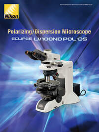
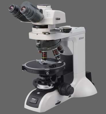
- Das LV100ND POL / DS verwendet Optiken mit hohen Extinktionsfaktoren, die die Erkennung und Beobachtung von hochpräzisen optischen Eigenschaftenermöglichen.
- Das LV100ND POL / DS ermöglicht die Analyse von Doppelbrechung, Extinktionswinkeln, Dehnungszeichen, Brechungsindizes, Doppelbrechungsgrößen und Pleochroismus von Asbest.
- Durch den Wechsel der Objektive und PH-Module können Asbestdispersionsfarben beobachtet werden, die den Brechungsindizes der Immersionsflüssigkeit entsprechen. Das LV100ND POL / DS ermöglicht sowohl die quantitative Analyse als auch die Beobachtung von Asbest durch Dispersionsfärbung.
- Ein vollständig drehbarer, kreisförmiger Messtisch bietet eine problemlose Probenrotation und eine genaue Analyse während der Polarisationsbeobachtung.
![](https://downloads.microscope.healthcare.nikon.com/production/imager/productphotos/2165/lv100nd-pol-ds_3cac538b21a254209ea06cf584646f29.jpg 1x, https://downloads.microscope.healthcare.nikon.com/production/imager/productphotos/2165/lv100nd-pol-ds_7fc2443c96341e0fab73dbc596e4b275.jpg 2x)
Hauptmerkmale
![](https://downloads.microscope.healthcare.nikon.com/production/imager/keyfeatures/LV100ND-POL-DS/2234/LV100ND-POL-Birefringence1_eab673dc52b0446b2b598a80abc7b050.jpg 1x, https://downloads.microscope.healthcare.nikon.com/production/imager/keyfeatures/LV100ND-POL-DS/2234/LV100ND-POL-Birefringence1_9b9989dbe1d056a5c35107959161a1c5.jpg 2x)
The cross Nicols, diagonal position
![](https://downloads.microscope.healthcare.nikon.com/production/imager/keyfeatures/LV100ND-POL-DS/2235/LV100ND-POL-Birefringence2_eab673dc52b0446b2b598a80abc7b050.jpg 1x, https://downloads.microscope.healthcare.nikon.com/production/imager/keyfeatures/LV100ND-POL-DS/2235/LV100ND-POL-Birefringence2_9b9989dbe1d056a5c35107959161a1c5.jpg 2x)
The cross Nicols, extinction position
![](https://downloads.microscope.healthcare.nikon.com/production/imager/keyfeatures/LV100ND-POL-DS/2238/LV100ND-POL-Extinction-angle1_eab673dc52b0446b2b598a80abc7b050.jpg 1x, https://downloads.microscope.healthcare.nikon.com/production/imager/keyfeatures/LV100ND-POL-DS/2238/LV100ND-POL-Extinction-angle1_9b9989dbe1d056a5c35107959161a1c5.jpg 2x)
The parallel Nicols, cleavage face aligned with the crosshair
![](https://downloads.microscope.healthcare.nikon.com/production/imager/keyfeatures/LV100ND-POL-DS/2237/LV100ND-POL-Extinction-angle2_eab673dc52b0446b2b598a80abc7b050.jpg 1x, https://downloads.microscope.healthcare.nikon.com/production/imager/keyfeatures/LV100ND-POL-DS/2237/LV100ND-POL-Extinction-angle2_9b9989dbe1d056a5c35107959161a1c5.jpg 2x)
The cross Nicols, extinction angle measured
![](https://downloads.microscope.healthcare.nikon.com/production/imager/keyfeatures/LV100ND-POL-DS/2241/LV100ND-POL-DS-elongation1_eab673dc52b0446b2b598a80abc7b050.jpg 1x, https://downloads.microscope.healthcare.nikon.com/production/imager/keyfeatures/LV100ND-POL-DS/2241/LV100ND-POL-DS-elongation1_9b9989dbe1d056a5c35107959161a1c5.jpg 2x)
Amosite Positive: Subtractive diagonal position
![](https://downloads.microscope.healthcare.nikon.com/production/imager/keyfeatures/LV100ND-POL-DS/2240/LV100ND-POL-DS-elongation2_eab673dc52b0446b2b598a80abc7b050.jpg 1x, https://downloads.microscope.healthcare.nikon.com/production/imager/keyfeatures/LV100ND-POL-DS/2240/LV100ND-POL-DS-elongation2_9b9989dbe1d056a5c35107959161a1c5.jpg 2x)
Crocidolite Negative: Additive diagonal position
![](https://downloads.microscope.healthcare.nikon.com/production/imager/keyfeatures/LV100ND-POL-DS/2247/LV100ND-POL-DS-refractive-index1_eab673dc52b0446b2b598a80abc7b050.jpg 1x, https://downloads.microscope.healthcare.nikon.com/production/imager/keyfeatures/LV100ND-POL-DS/2247/LV100ND-POL-DS-refractive-index1_9b9989dbe1d056a5c35107959161a1c5.jpg 2x)
Der obere Teil einer Probe ist fokussiert
![](https://downloads.microscope.healthcare.nikon.com/production/imager/keyfeatures/LV100ND-POL-DS/2246/LV100ND-POL-DS-refractive-index2_eab673dc52b0446b2b598a80abc7b050.jpg 1x, https://downloads.microscope.healthcare.nikon.com/production/imager/keyfeatures/LV100ND-POL-DS/2246/LV100ND-POL-DS-refractive-index2_9b9989dbe1d056a5c35107959161a1c5.jpg 2x)
Der untere Teil einer Probe ist fokussiert
![](https://downloads.microscope.healthcare.nikon.com/production/imager/keyfeatures/LV100ND-POL-DS/2251/LV100ND-POL-DS-birefringence-magnitude1_5aee3c2798fab066547b202a83b201a8.jpg 1x, https://downloads.microscope.healthcare.nikon.com/production/imager/keyfeatures/LV100ND-POL-DS/2251/LV100ND-POL-DS-birefringence-magnitude1_b378458833c32c493443ede68db69be0.jpg 2x)
Diagonale Position
![](https://downloads.microscope.healthcare.nikon.com/production/imager/keyfeatures/LV100ND-POL-DS/2250/LV100ND-POL-DS-birefringence-magnitude2_5aee3c2798fab066547b202a83b201a8.jpg 1x, https://downloads.microscope.healthcare.nikon.com/production/imager/keyfeatures/LV100ND-POL-DS/2250/LV100ND-POL-DS-birefringence-magnitude2_b378458833c32c493443ede68db69be0.jpg 2x)
Subtraktive Diagonalposition bestätigt (eine Testplatte wird eingefügt)
![](https://downloads.microscope.healthcare.nikon.com/production/imager/keyfeatures/LV100ND-POL-DS/2249/LV100ND-POL-DS-birefringence-magnitude3_5aee3c2798fab066547b202a83b201a8.jpg 1x, https://downloads.microscope.healthcare.nikon.com/production/imager/keyfeatures/LV100ND-POL-DS/2249/LV100ND-POL-DS-birefringence-magnitude3_b378458833c32c493443ede68db69be0.jpg 2x)
Analysator Rotationswinkel gemessen (Senarmont Kompensator wird eingesetzt)
![](https://downloads.microscope.healthcare.nikon.com/production/imager/keyfeatures/LV100ND-POL-DS/2255/LV100ND-POL-DS-Pleochroism1_eab673dc52b0446b2b598a80abc7b050.jpg 1x, https://downloads.microscope.healthcare.nikon.com/production/imager/keyfeatures/LV100ND-POL-DS/2255/LV100ND-POL-DS-Pleochroism1_9b9989dbe1d056a5c35107959161a1c5.jpg 2x)
Parallel zur Schwingungsrichtung des Polarisators
![](https://downloads.microscope.healthcare.nikon.com/production/imager/keyfeatures/LV100ND-POL-DS/2256/LV100ND-POL-DS-Pleochroism2_eab673dc52b0446b2b598a80abc7b050.jpg 1x, https://downloads.microscope.healthcare.nikon.com/production/imager/keyfeatures/LV100ND-POL-DS/2256/LV100ND-POL-DS-Pleochroism2_9b9989dbe1d056a5c35107959161a1c5.jpg 2x)
Orthogonal zur Schwingungsrichtung des Polarisators