- ko Change Region
- Global Site
Application Notes

Cell cycle analysis of keratinocytes and label-free cell counting of mitotic cells using Volume Contrast
2022 년 03 월
Keratinocytes are epidermal cells used in various studies such as cosmetics development, drug discovery, and regenerative medicine. In keratinocytes, autophagy induction is known to regulate melanosome degradation, which is being used to study whitening ingredients in cosmetics development. Psoriasis, an autoimmune disease, is known as a skin disease caused by abnormal cell proliferation of keratinocytes.
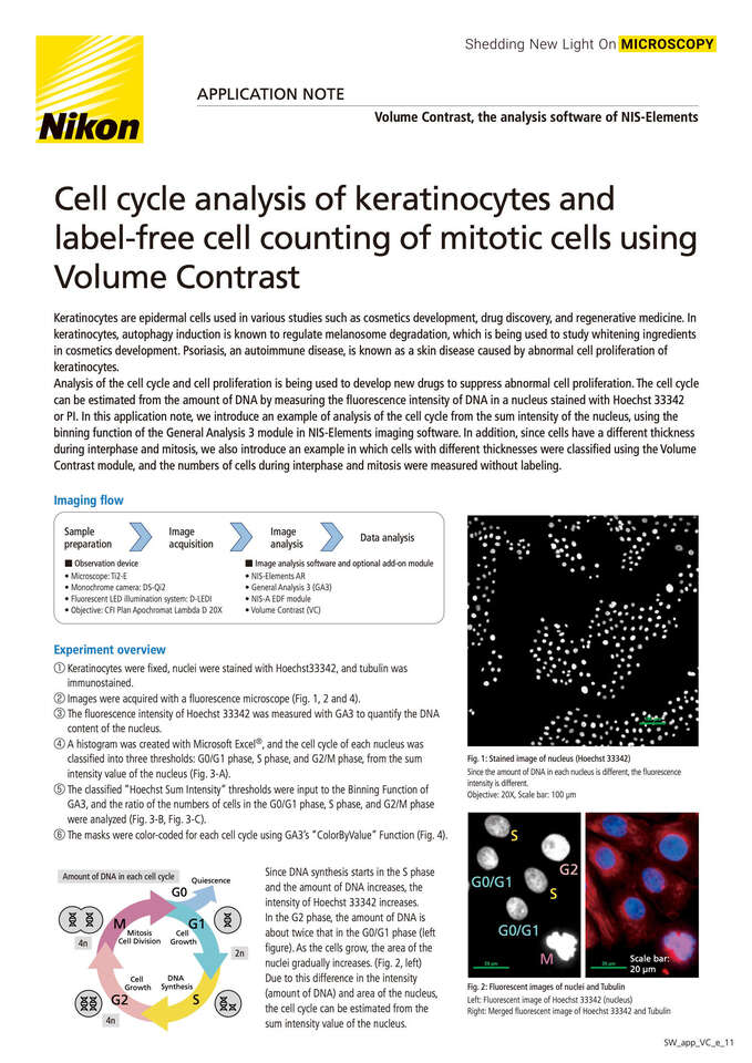
Analysis of the cell cycle and cell proliferation is being used to develop new drugs to suppress abnormal cell proliferation. The cell cycle can be estimated from the amount of DNA by measuring the fluorescence intensity of DNA in a nucleus stained with Hoechst 33342 or PI. In this application note, we introduce an example of analysis of the cell cycle from the sum intensity of the nucleus, using the binning function of the General Analysis 3 module in NIS-Elements imaging software. In addition, since cells have a different thickness during interphase and mitosis, we also introduce an example in which cells with different thicknesses were classified using the Volume Contrast module, and the numbers of cells during interphase and mitosis were measured without labeling.
