니콘인스트루먼트코리아 | Korea
- ko Change Region
- Global Site

2021 년 06 월
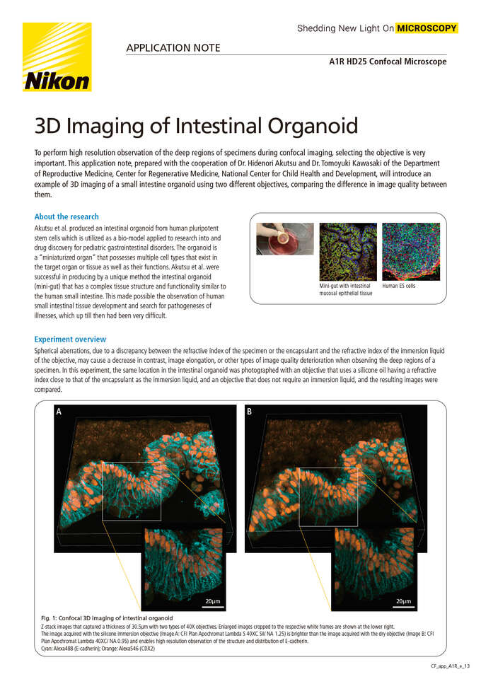
To perform high resolution observation of the deep regions of specimens during confocal imaging, selecting the objective is very important. This application note, prepared with the cooperation of Dr. Hidenori Akutsu and Dr. Tomoyuki Kawasaki of the Department of Reproductive Medicine, Center for Regenerative Medicine, National Center for Child Health and Development, will introduce an example of 3D imaging of a small intestine organoid using two different objectives, comparing the difference in image quality between them.