 ®
®
Nikon Instruments Inc. | Americas
- es Change Region
- Global Site
Centro de excelencia
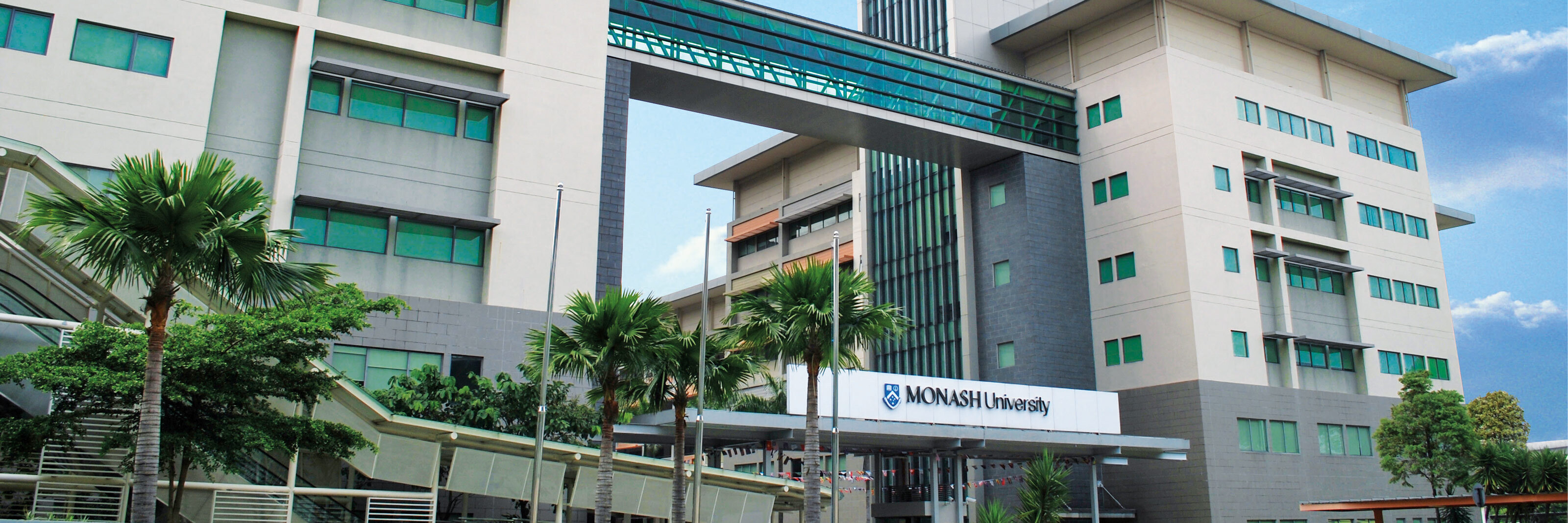
The Monash University Malaysia Optical Imaging Platform (MUM OIP) has partnered with Nikon, a global leader in optical imaging technology, to establish the Nikon Center of Excellence (NCoE) at Monash University Malaysia. The NCoE strengthens our imaging capabilities and provides researchers with access to advanced microscopy technologies, technical support and training.
Beyond providing access to instruments, the NCoE aims to support researchers through microscopy training, application guidance, image analysis and consultation. We also hope to build stronger connections with researchers, industry partners and the wider bioimaging community in Malaysia and Southeast Asia.
The MUM OIP-Nikon Center of Excellence is a place to explore new imaging possibilities, learn microscopy, and support researchers in getting the most out of their imaging experiments.
email hidden; JavaScript is required
The Nikon AX R NSPARC is Nikon’s latest flagship confocal microscope, designed for high-speed, high-resolution, and gentle imaging of both live and fixed specimens. It features 2K resonant scanning with an expanded 25 mm field of view (FOV) for rapid imaging with reduced photobleaching and phototoxicity, making it ideal for sensitive live-cell applications. The integrated NSPARC Image Scanning detector uses a highly sensitive APD detector array to deliver enhanced signal-to-noise ratio, deep tissue imaging, and super-resolution imaging of fine cellular and subcellular structures.
Built on Nikon’s flagship ECLIPSE Ti2 inverted microscope platform, this advanced live-cell imaging system is designed to capture dynamic cellular processes under physiologically controlled conditions. Fully integrated with NIS-Elements, it supports automated time-lapse imaging, multi-position acquisition, and high-content live-cell experiments. The system also features 2D/3D Deconvolution for improved image clarity, JOBS for automated imaging workflows, and General Analysis 3 (GA3) for quantitative analysis of cell migration, proliferation, viability, morphology, and other live-cell assays.
Stereo microscope with 25X zoom range, suitable for fluorescence imaging, developmental studies, live and fixed cells.


