Nikon Corporation Healthcare Business Unit | Asia, Oceania & Middle East
- en Change Region
- Global Site

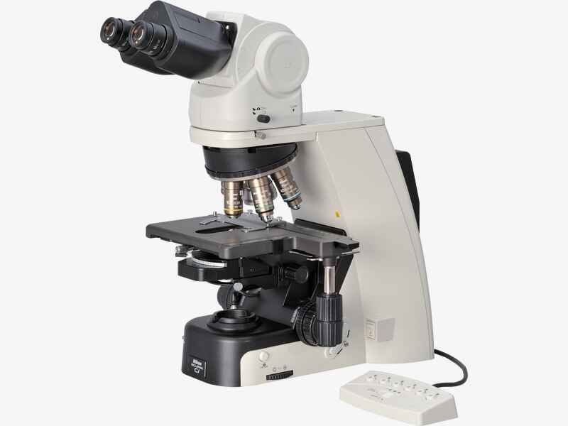
To meet the demands of clinical laboratory specialists and researchers, Nikon has reviewed all aspects of microscope usability to develop the ECLIPSE Ci series of microscopes, which combines superior functionality with operational ease. The Ci is designed to ensure natural posture while viewing images, sample changing and capturing images. Bright, eco-friendly LED illumination reduces the need for frequent lamp replacement*. A variety of accessories are available that support various imaging techniques.
* Halogen model (Ci-S) is also available
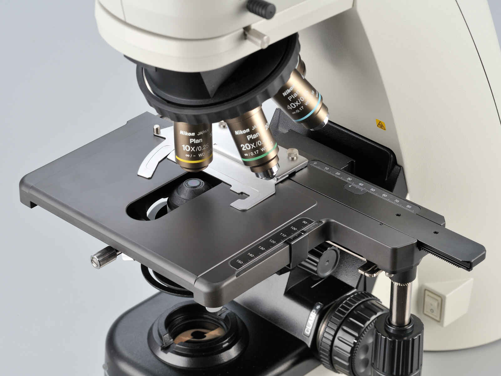
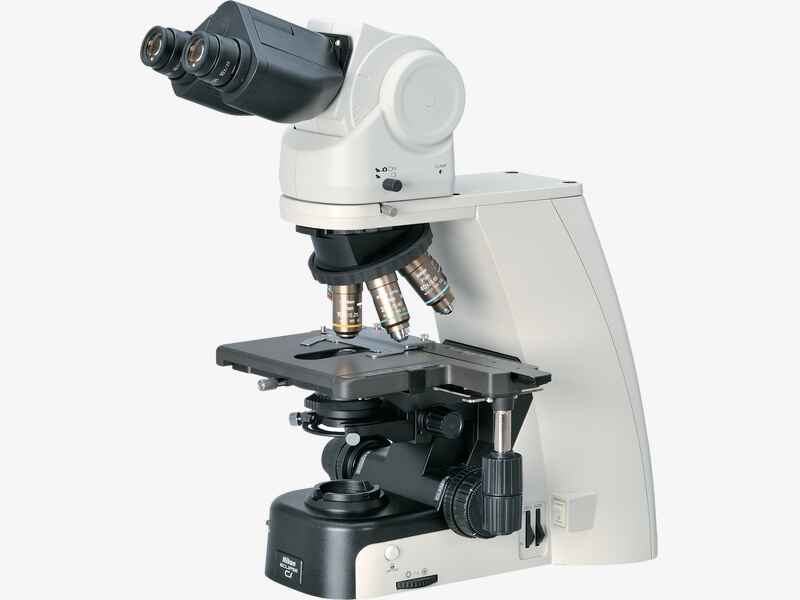
Equipped with motorized nosepiece and automatic intensity recall, the Ci-E is ideal for applications that require frequent magnification switching.
Suitable for applications requiring a traditional halogen light source.
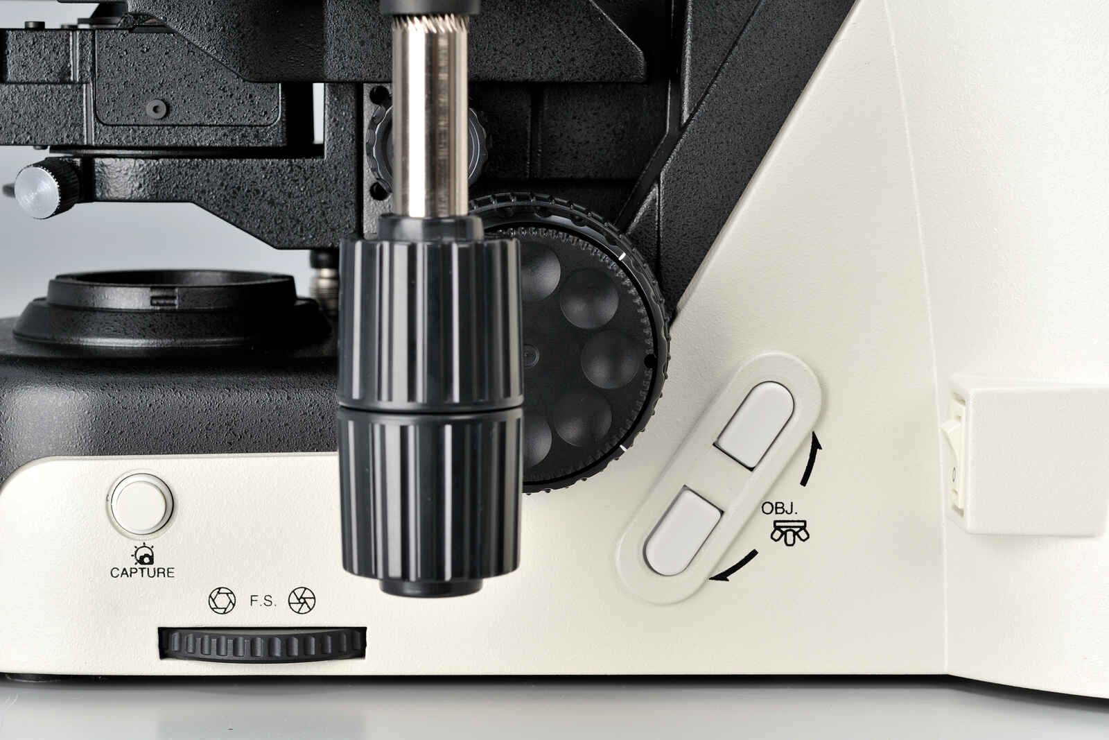
Magnification can be easily changed during observation with the touch of a button on the ECLIPSE Ci-E microscope body. The buttons can also be programmed for specific objective lenses for easy switching between specific pairs. User-defined light intensity for each magnification is automatically saved and reproduced when magnification is changed. The remote control pad can also be used to easily change magnifications.
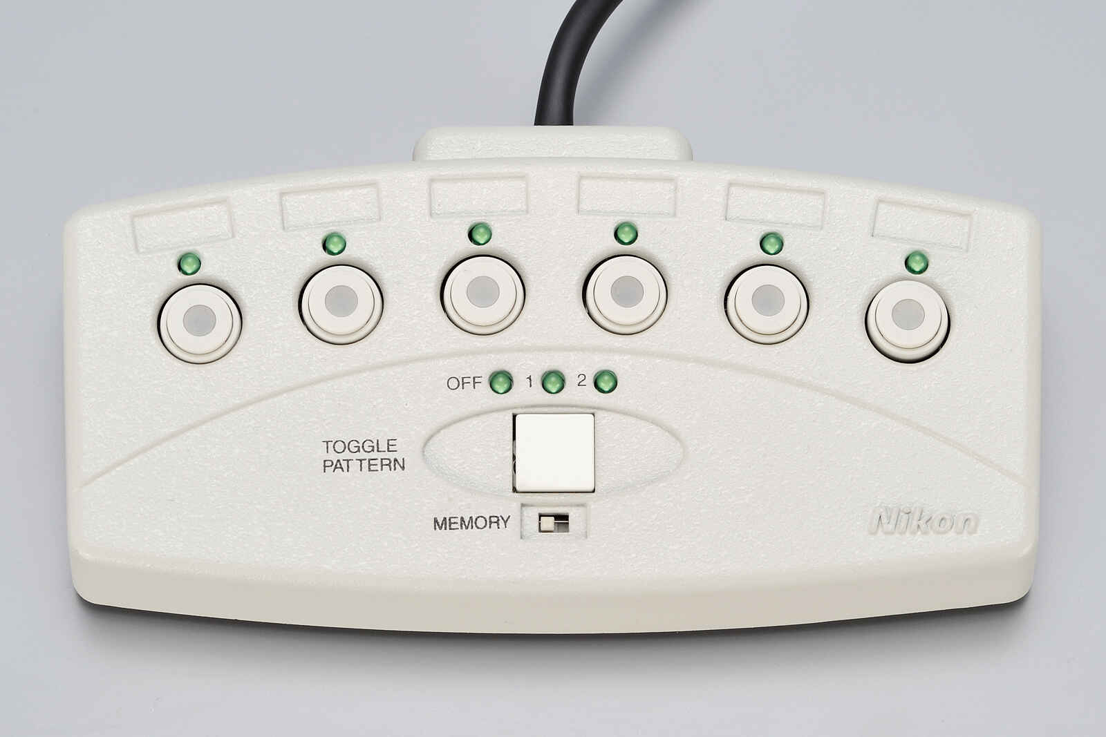
Nosepiece rotation buttons
Remote control pad
The Eco-illumination* is a low-power-consuming, eco-friendly illumination system that produces uniform brightness and reduces the cost and effort of lamp replacement, thanks to its 60,000 hour, high-luminescent LED. By combining a collimator lens, fly-eye optics and LED illumination, bright and edge-to-edge uniform images can be obtained even at high magnifications. The LED illuminator features low-heat generation and provides the same color temperature at every magnification.
* Halogen model (Ci-S) is also available

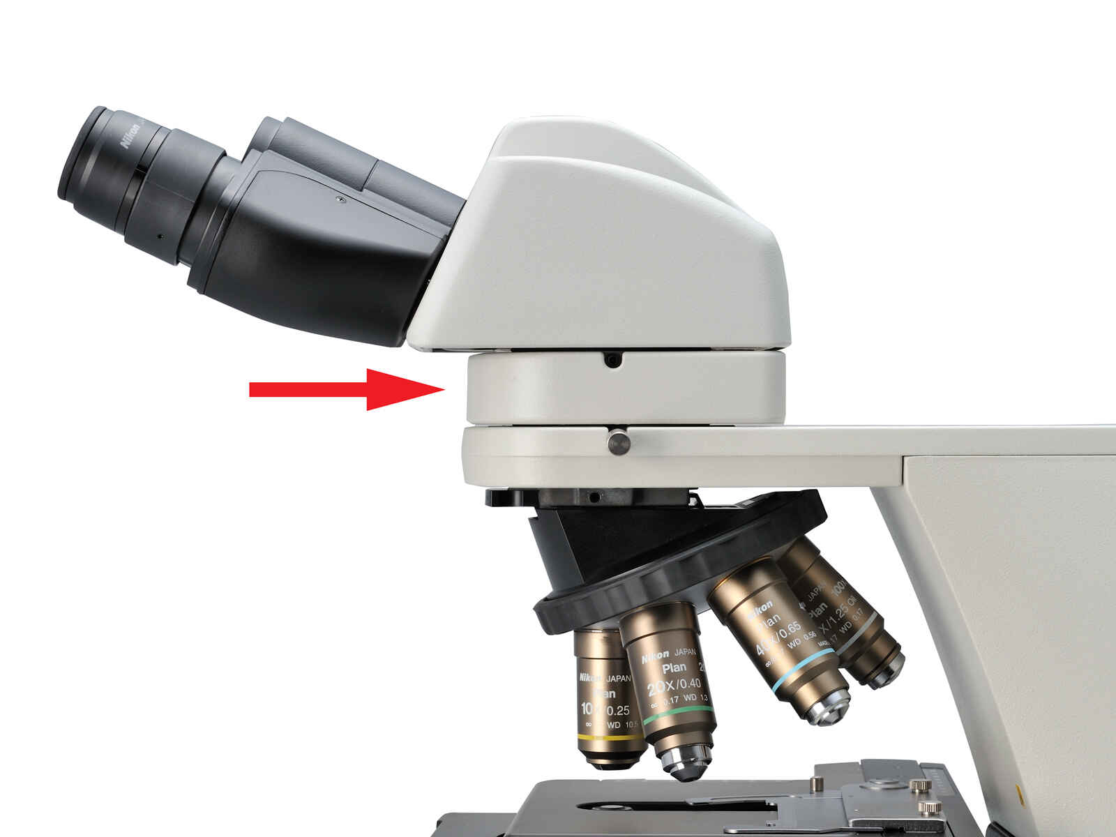
Using the ergonomic binocular tube, which features an eyepiece that can be inclined from 10° to 30°and extended up to 40 mm, the microscope can be adjusted for natural posture. The eyelevel riser lifts the eyepiece tube in 25 mm increments (up to 100 mm*) and can easily be adapted to suit users with different eye-point heights.
* Up to 50 mm with ergonomic binocular tube.
Ergonomic binocular tube
Eyelevel riser
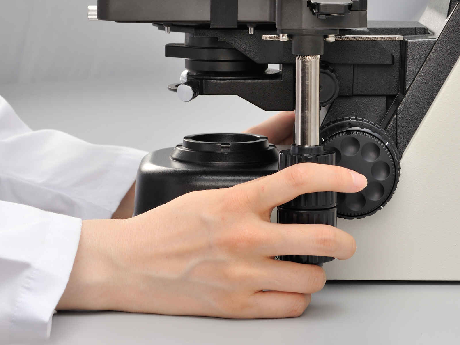
With the addition of a nosepiece spacer, the stage height can be lowered 20 mm from the standard position, reducing strain during frequent specimen change. The stage handle height can also be changed to ensure a comfortable hand position. The stage height can be locked using the refocusing knob, allowing quick refocusing after specimen changes. The stage is coated with a high-durability, scratch-resistant ceramic coating.
Without spacer (left), with spacer (right)
Height adjustable stage handle
Ceramic-coated stage
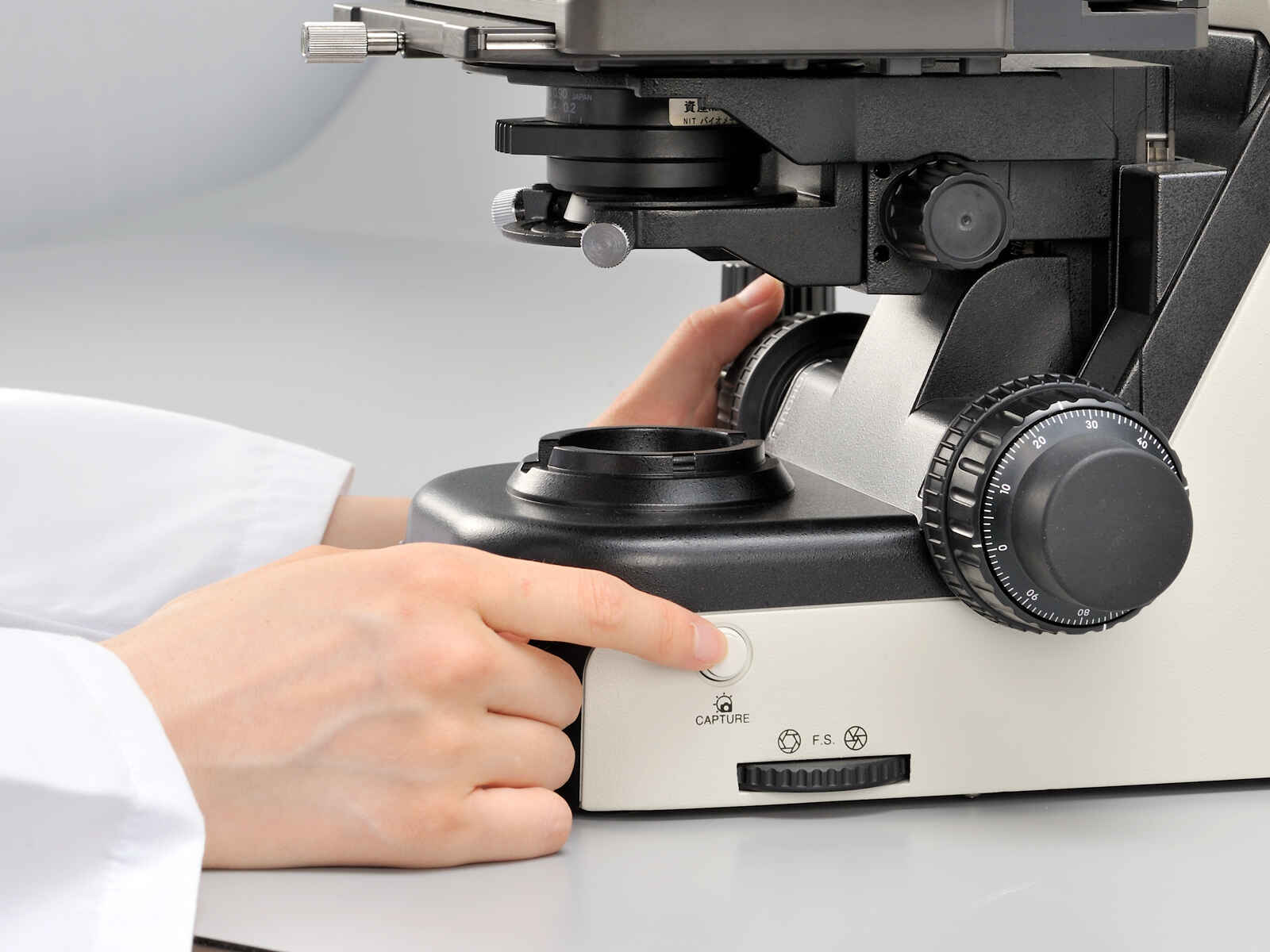
While maintaining your observation posture, simply press the capture switch to easily acquire images with the Digital Sight series microscope cameras.
The free software package NIS-Elements LE features a Scene Mode that automatically sets optimal imaging conditions for each observation method. In addition, its streaming function allows you to share images with a remote PC.
* NIS-Elements LE is not for clinical diagnostic use.
Image capture button
NIS-Elements LE camera control software for tablet PC
Digital pathology via network
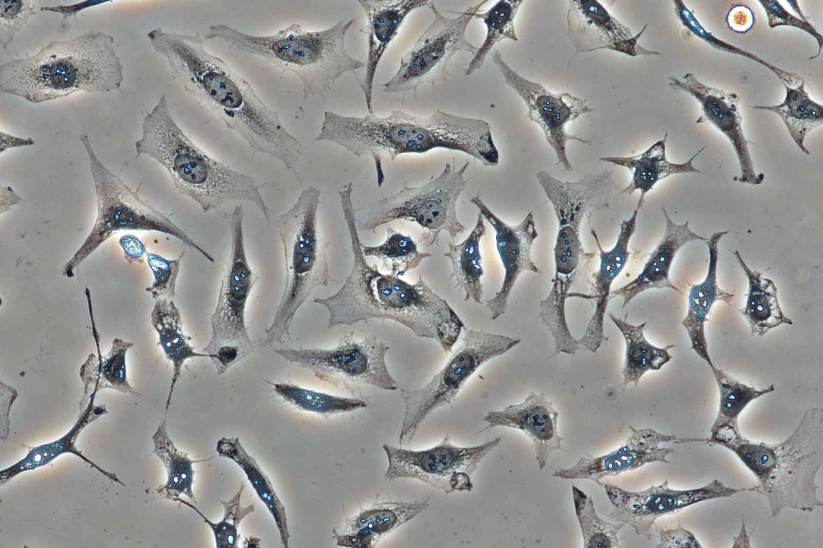
High-contrast images with neutral background coloration regardless of the magnification range can be captured. This observation technique is suitable for observation of unstained structures.
Phase contrast accessories and objective lenses
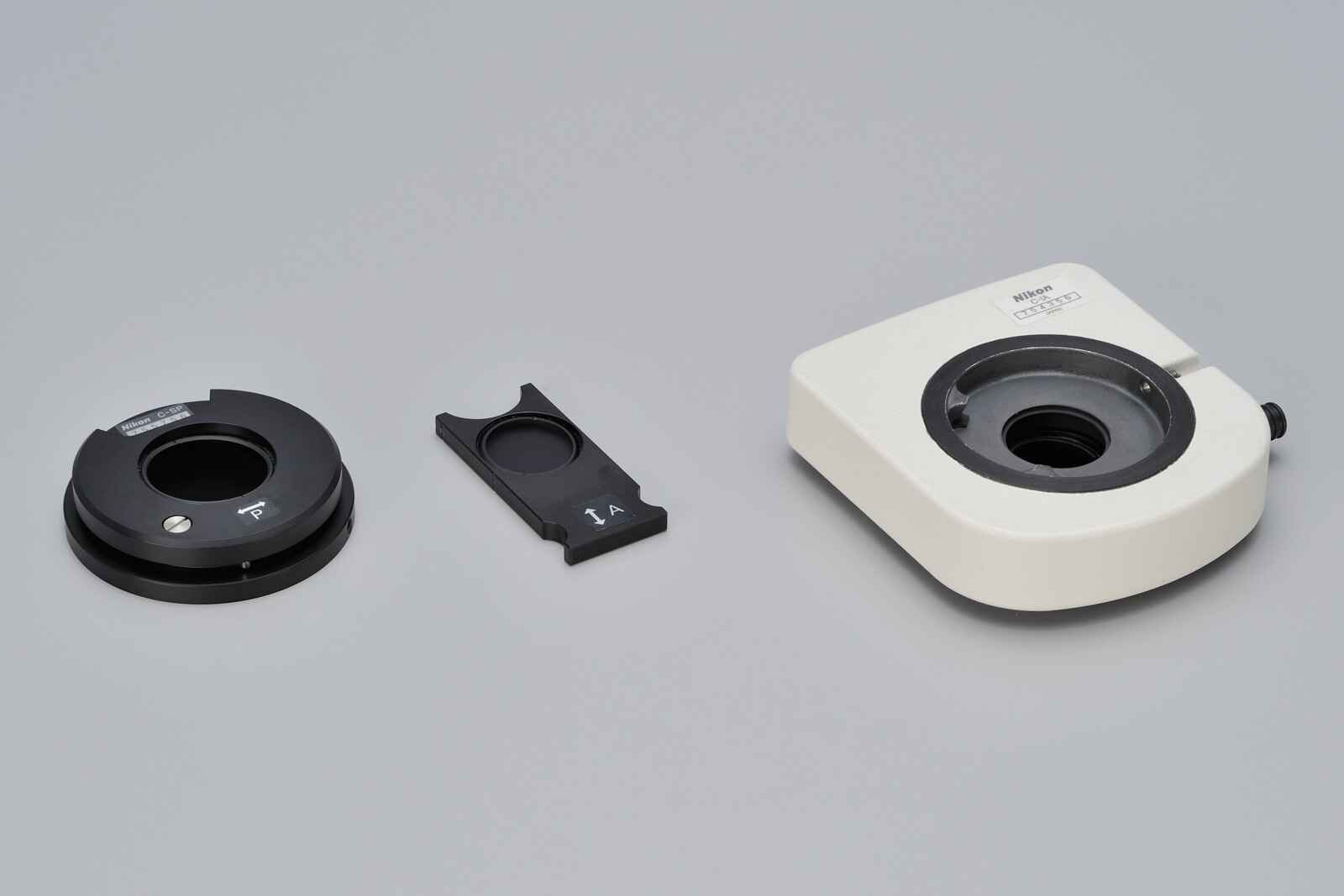
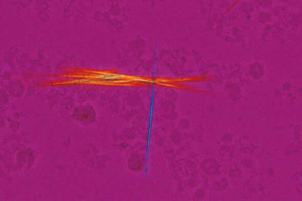
Ideal for observing bi-refringent samples such as collagen, amyloids and crystals.
* Two types of analyzer are available: intermediate tube type and nosepiece slider type.
2.8-Dihydroxyadenine crystals, Simple polarizing, CFI Plan Fluor 40X
Department of Clinical laboratory, Nihon University Itabashi Hospital
Simple polarizing accessories
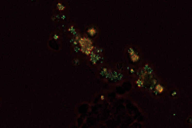
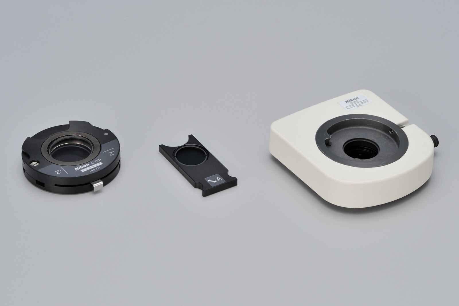
Enables Identification of uric acid crystals by changes in the interference color. This technique is ideal for gout and pseudo-gout tests.
* Two types of analyzer are available: intermediate tube type and nosepiece slider type.
Sodium urate crystals, Sensitive color polarizing, CFI Plan Fluor 40X
Department of Clinical laboratory, Nihon University Itabashi Hospital
Sensitive color polarizing accessories
Enables clear observation of blood or minute structures such as flagella. Dry- and oil-type condensers are available. An expander lens is utilized for brighter imaging.
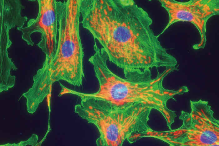
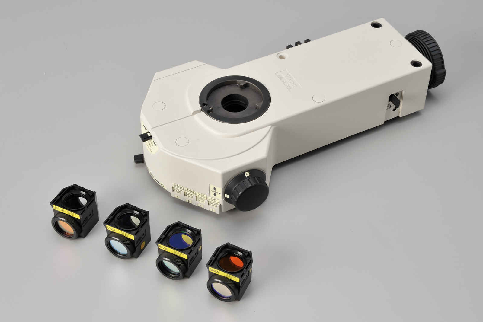
Compact epi-fluorescence attachments that utilize noise terminating mechanisms allow weakly fluorescing specimens to be captured with great clarity and brightness. Both CI-FL epi-fluorescence attachment (incorporates up to 4 filter cubes) and D-FL epi-fluorescence attachment (incorporates up to 6 filter cubes) allow easy switching of filter cubes. High-optical-performance objective lenses for epi-fluorescence imaging, including the CFI Plan Apochromat Lambda series and the CFI Plan Fluor series, are available.
CI-FL epi-fluorescence attachment and filter cubes