
Compact, high-definition, high-speed and high-sensitivity C-mount camera.
DS-Fi3 is a high-definition color microscope camera equipped with a 5.9 megapixel CMOS image sensor. Its high-speed data readout, superior color reproduction and high quantum efficiency are optimal for imaging in various observations, such as brightfield, DIC, phase contrast and fluorescence observation.
* DS-Fi3 is not for clinical diagnostic use.
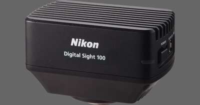
Key Features
High definition imaging
DS-Fi3 is equipped with a 5.9 megapixel CMOS image sensor, which enables the capture of high-definition images of up to 2880 x 2048 pixels. With this new CMOS image sensor and high-speed data transfer via USB 3.0, DS-Fi3 enables fast focusing even in high-resolution imaging, and efficient image acquisition when using a wide range of illumination techniques.

Tubular adenoma, HE staining (Objective: CFI Plan Apochromat Lambda 4X)
Photos courtesy of: Dr. Yasunori Ohta, Department of Pathology, IMSUT Hospital, Institute of Medical Science, The University of Tokyo
High sensitivity and low noise
The DS-Fi3 has significantly higher quantum efficiency and lower readout noise than conventional models. It allows for the capture of brighter images with higher S/N ratios in fluorescence observation modes.

Breast cancer, FISH method (Objective: CFI Plan Apochromat Lambda 100X Oil)
Photos courtesy of: Hironao Kusakari, Diagnostic Pathology, St. Marianna University Hospital
High-speed live display
Due to the high-speed data readout by the CMOS image sensor and high-speed USB3.0 data transfer, the DS-Fi3 displays live images of full 2880 x 2048-pixel images at 15 fps, and 1440 x 1024-pixel images at 30 fps. This enables smooth display during focusing and the selection of observation locations.
Excellent color reproducibility
Nikon is well known for outstanding and lifelike color reproduction, and developing superior algorithms for creating results that look like the actual samples. These algorithms are used in all of the color cameras in the digital sight lineup.

Uterine cervix Pap staining (Objective: CFI Plan Apochromat Lambda 40XC)
Photos courtesy of: Kazuhiro Mita, Department of Pathology, Yokohama City University Hospital

Bone marrow (Objective: CFI Plan Achromat NCG 40X)
Photos courtesy of: Clinical Laboratory Department, Yokohama City University Hospital
Direct connection to a PC
The DS-Fi3 allows direct connection to a PC via a USB3.0 interface to control the camera and capture images using NIS-Elements imaging software.