Nikon Corporation Healthcare Business Unit | Asia, Oceania & Middle East
- en Change Region
- Global Site

Nikon Imaging Center
The Nikon Imaging Center at Northwestern University Feinberg School of Medicine (NIC@NU-FSM) is an integral component of the Northwestern University Cell Imaging Facility, supported by Nikon, the Feinberg School of Medicine, Department of Cell & Molecular Biology and the Robert H. Lurie Comprehensive Cancer Center.
Established in 2008, the NIC@NU-FSM carries an important mission to:
email hidden; JavaScript is required
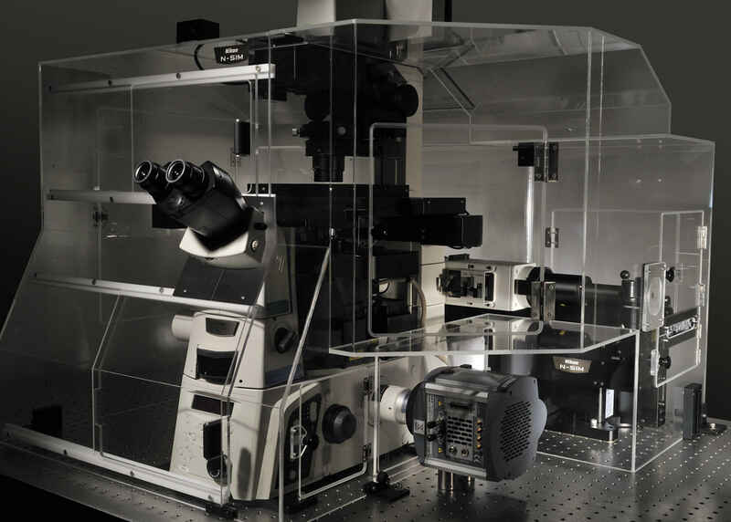
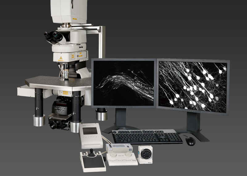
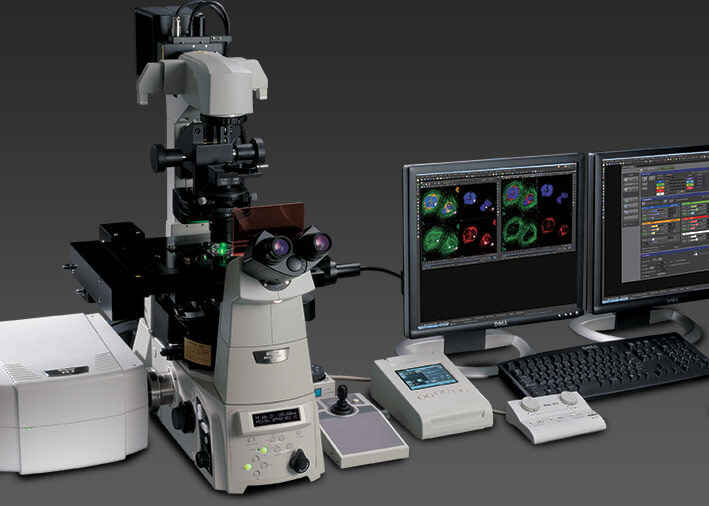
Featuring both Nikon's N-SIM structured illumination system and incubation, this system provides a powerful platform for live-cell super-resolution imaging.
NIS-Elements software
The BioStation IM-Q incorporates a microscope, an incubator and a high-sensitivity cooled CCD camera in a compact body. This all-in-one package provides a stable environment for live cells and advanced solutions for simple long-term time-lapse data acquisition.
The BioStation IM-Q incorporates a microscope, an incubator and a high-sensitivity cooled CCD camera in a compact body. This all-in-one package provides a stable environment for live cells and advanced solutions for simple long-term time-lapse data acquisition.
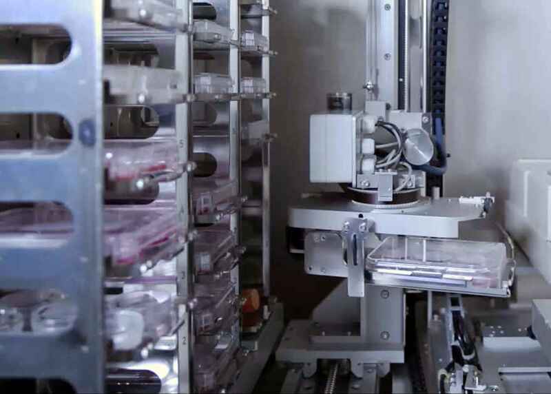
The BioStation CT combines the cell culture and imaging environments to enable accurate monitoring of individual cells and colonies while maintaining stable environmental conditions.
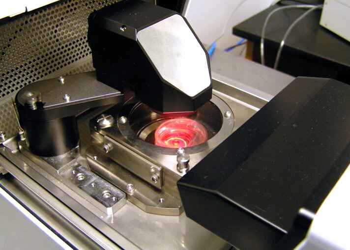
The AZ/C2 is a macro confocal microscope system which allows for the capture of confocal images allowing for better resolution of 3D specimens.
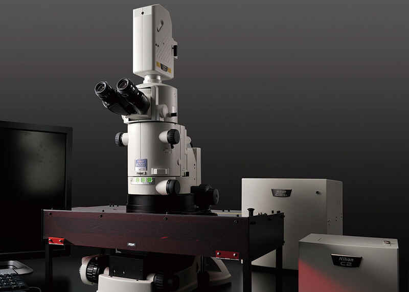
C2 point scanning confocal system
LUN-4 4-line laser unit (408nm, 488nm, 561nm, 640nm)
NIS-Elements software
Deep tissue imaging is made possible using this Nikon A1R MP multiphoton system configured on Ni-E upright microscope.
Ni-E upright microscope
NIS-Elements software
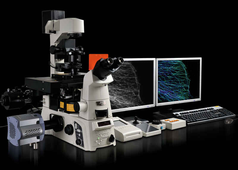
This multimodal TIRF, super-resolution N-STORM, and spinning disk confocal system is adaptable to a variety of samples.
N-STORM super-resolution single molecule localization microscopy system
Yokogawa CSU-X1 spinning disk confocal
NIS-Elements software
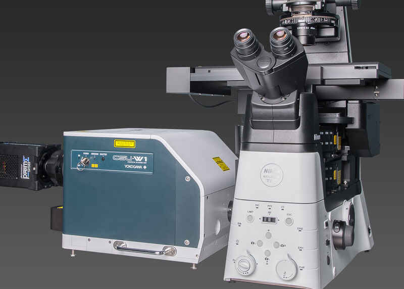
Combining the Nikon Ti2-E microscope with Yokogawa CSU-W1 spinning disk and emission splitting optics unlocks fast and large field of view confocal imaging.
Ti2-E inverted microscope with Perfect Focus System 4 (PFS4)
Yokogawa CSU-W1 spinning disk confocal
NIS-Elements software
This fast and large field of view widefield system features a Nikon Ti2-E and Lumencor Spectra III light engine.
Ti2-E inverted microscope with Perfect Focus System 4 (PFS4)
Large format DS-Qi2 monochrome CMOS camera
NIS-Elements software
Using the Nikon A1Rsi resonant scanning confocal system with spectral detector, this system is made for linear unmixing of multiplynlabeled samples.
NIS-Elements software