


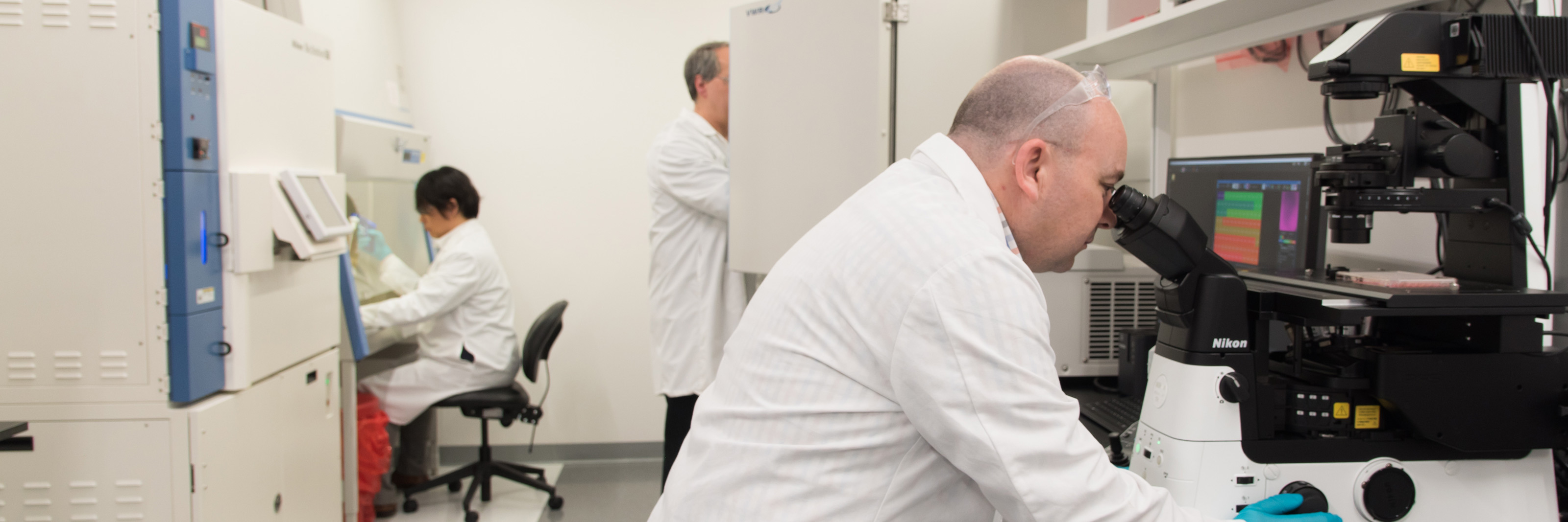
Applications & Technology
Provided here is an overview of the various applications supported by the Nikon BioImaging Lab, detailing imaging, analysis, and other tools we have at our disposal. Click to jump to a specific application, or scroll down to start exploring.
High Content Imaging and Analysis
Technology: BioPipeline LIVE, A1R HD25 Confocal, NIS.ai deep learning software, microfluidics, cell culturing services
High content imaging is about imaging more – more detection channels, more time points, more data. It’s no wonder that high content imaging and analysis is a powerful tool for so many applications, such as phenotypic screening in drug discovery research. However, the greatest strength of high content imaging – the volume of rich data created – can also be its greatest drawback, creating critical bottlenecks in experiment design, data storage, and image analysis. At the Nikon BioImaging Lab, our high content imaging services are centered around the BioPipeline Live, a stable yet flexible platform for long-term live-cell high content that addresses common process bottlenecks to provide 24/7 operational efficiency.
High content imaging with the BioPipeline LIVE is great for applications ranging from cell health monitoring, to toxicology studies, and much more. For imaging thicker samples, our BioPipeline Live system features a Nikon A1R HD25 resonant scanning confocal system for acquiring data deep in samples such as spheroids, organoids, organ-on-a-chip, and tissue. This system also features our NIS.ai deep learning software modules, which can be trained to perform a number of different analysis tasks, such as segmentation, and can be integrated into an automated imaging and analysis pipeline. Microfluidics-based cell manipulation and cell culturing services are also available.

3D Imaging
Technology: BioPipeline LIVE, A1R HD25 Confocal, Ti2-E Inverted Microscope
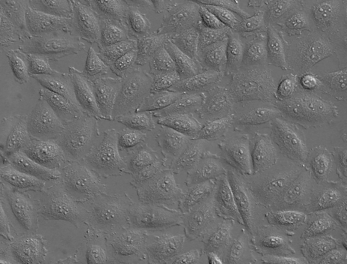
Cell biology and other fields have long relied on in vitro imaging of a single layer of cultured cells adhering to a flat surface, but the limitations of such model systems are being increasingly recognized, and more physiologically-relevant models are gaining in popularity, including spheroids, organoids, organ-on-a-chip, and similar systems. While these systems provide better models of organismal biology, they have non-trivial thickness and 3D structure, which limits the application of routine 2D widefield imaging approaches.
Our BioPipeline LIVE system features an A1R HD25 confocal, our flagship point-scanning confocal system for deep imaging tens of micrometers or more into the sample. Our Ti2-E inverted microscope is also equipped for 3D widefield imaging, featuring integrated deconvolution software for achieving optical sectioning in samples to approximately 20 micrometers thick. While 3D imaging by widefield deconvolution doesn’t provide the instantaneous 3D sectioning of a confocal instrument, it is fast and often the superior choice for low-signal samples. We understand the nuances that inform proper choice of 3D imaging technique, and are here to make sure you are successful.
Pilha Z de um organoide renal adquirido usando um sistema confocal Nikon A1R HD25.
Vermelho: Phalloidin-Alexa Fluor 568 (actina filamentosa)
Azul: DNA (núcleos)
Fluorescence Imaging
Technology: BioPipeline LIVE, A1R HD25 Confocal, BioStation CT, Ti2-E Inverted Microscope
Fluorescence microscopy is widely considered to be one of the most sensitive imaging methods for detecting a given molecular target with high specificity. Fixed samples are generally labeled with dye-conjugated antibodies using standard immunohistochemistry/immunofluorescence techniques. Live samples can be made to genetically express a fluorescent protein fused to a target protein of interest or stained with membrane-permeable organic dyes.
Fluorescence underpins a number of important imaging methods, providing a convenient mechanism for multichannel imaging via the labeling of different targets with spectrally-distinct fluorophores. All of our systems are built to perform fluorescence imaging, however there are many factors that must be considered to do so successfully. At the Nikon BioImaging Lab, we have an intimate understanding of concerns related to phototoxicity, sample preparation (including fluorophore selection), and other such critical factors that help determine the success of a fluorescence-based experiment.
The A1R HD25 features laser illumination, and the BioStation CT and Ti2-E inverted microscope feature LED illumination. Specifically, our Ti2-E features a fast color-switching Lumencor Spectra-X 7-channel LED light engine for advanced multiplexing experiments. The A1R HD25 is configured with 6 different color laser lines, including 445 nm and 514 nm lines, which are commonly used for CFP/YFP FRET studies. It is also capable of performing targeted FRAP and photostimulation experiments. We stand ready with the expertise to craft successful fluorescence-based assays, having a deep knowledge of fluorophores, microscope settings, mounting media, and related concerns.
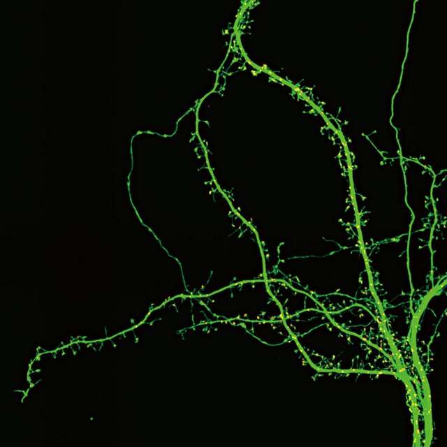
Neurônio do hipocampo de um camundongo. Verde: GFP (dendritos), vermelho: PSD-95-TagRFP (marcador pós-sináptico). Imagem capturada usando uma objetiva Nikon CFI SR HP Apochromat TIRF 100XC Oil, cortesia dos: Drs. Yutaro Kashiwagi e Shigeo Okabe, Departamento de Neurobiologia Celular, Escola de Graduação em Medicina e Faculdade de Medicina da Universidade de Tóquio.
High-Resolution Imaging
Technology: A1R HD25 Confocal, Ti2-E Inverted Microscope
It’s easy to find a microscope providing high magnification, but it takes much more than magnification alone to acquire quality high-resolution images. Our A1R HD25 confocal system provides both optical sectioning – the ability to resolve a distinct 2D section in a 3D sample – and high-resolution imaging of sample features in that 2D section. To maximize the resolution of our confocal instrument, we have the A1-ER enhanced-resolution software module for deconvolution of A1R HD25 data, which includes detailed point-spread function (PSF) models for Nikon’s market-leading objective lenses.
The Ti2-E inverted microscope is also capable of high-resolution multi-dimensional imaging – able to acquire data in 3D, over time, and in multiple color channels similar to the A1R HD25 – and includes deconvolution software for maximizing resolution and optical sectioning.
It’s not just the microscope that goes into making high resolution images, equally important is proper sample preparation. If, for example, your sample is mounted in a medium with the wrong refractive index, optical resolution will suffer significantly, especially in the Z direction. At the Nikon BioImaging Lab we have you covered on both fronts, with expertise in both high-resolution microscopy and sample preparation.

Visualizações XY e XZ dos cílios cocleares, antes (esquerda) e depois (direita) da deconvolução, usando o módulo de resolução aprimorada A1-ER. Dados de imagem adquiridos usando um Nikon A1R com diâmetro de orifício de 1 unidade Airy.
Color Imaging
Technology: Ti2-E Inverted Microscope
Our Ti2-E inverted microscope is equipped with a Lumencor LIDA light source for transmitted light (diascopic) illumination. The LIDA consists of individual red, green, and blue LEDs that can be triggered in succession, imaging each color sequentially, and combining the three images into a composite RGB color image. This is an advantage as it allows us to avoid using single exposure color cameras, which have lower sensitivity, spatial resolution, and dynamic range than their monochrome counterparts. Furthermore, the intensity of each LED color is individually addressable, unlocking very fine hardware-based control of color balancing without image post-processing or having to move any system components.
This technology is beneficial not only for pathologists and histologists, but to anyone looking for a modern approach to quantitative color imaging. This is even beneficial for live cell imaging applications as the LIDA does not feature any UV or near-IR light in its spectrum. In addition to brightfield, other techniques we support that can benefit from color imaging include polarized light, differential interference contrast (DIC), and darkfield microscopies. As always, the Nikon BioImaging Lab staff is here to advise you on best practices for your entire microscopy imaging and analysis workflow, including color imaging.
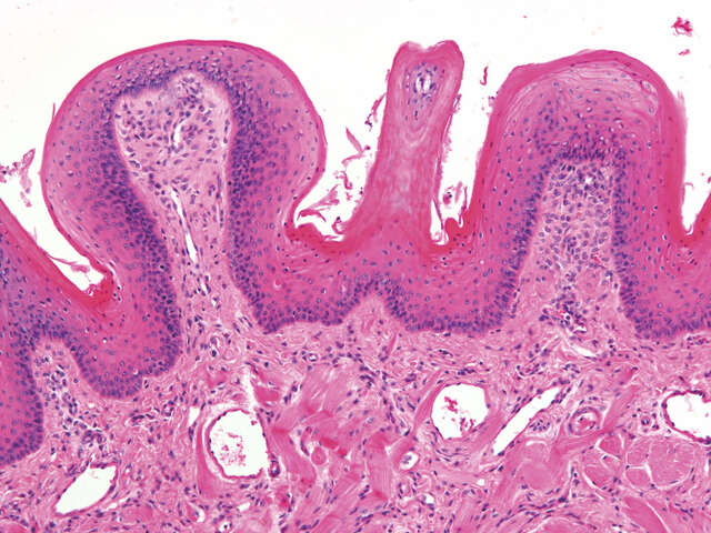
Brightfield color image of an H&E stained mammalian skeletal muscle tissue section acquired using a Nikon CFI Plan Apo Lambda 10X objective.
Label-free Imaging
Technology: BioPipeline LIVE, BioStation CT, BioStudio-T, ECLIPSE Ti2-E, NIS.ai
While fluorescence imaging might be one of the most popular contrasting techniques for optical microscopy, it is also one of the most invasive. Fluorophores are prone to photobleaching, limiting their useful lifetime, and resulting in the need for greater and greater illumination intensity to excite a similar amount of emission as the experiment progresses. This difficulty is compounded by the phototoxic response of live cells to high-intensity illumination. Assays using sensitive cell types, such as stem cells, may not be completely compatible with fluorescence imaging of the desired target.
Cell health concerns are helping to fuel the renewed popularity of label-free imaging techniques, which help avoid both the need for high-intensity illumination as well as perturbation of the system by the labeling process.
Artificial intelligence-based machine learning and deep learning analysis techniques are also helping to enable this trend. For example, the Convert.ai module (one of several NIS.ai deep learning software modules) automates the identification of key cell features from differential interference contrast (DIC) data, such as the simulated DAPI staining in the figure here. All of our systems support label-free imaging, with available techniques including new volume contrast, phase contrast, differential interference contrast (DIC), darkfield, and brightfield. Contact us today to see if label-free imaging will work for you.
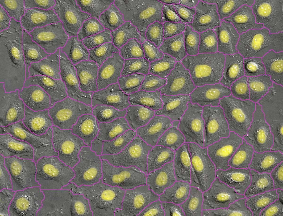 Convert.ai
 Original
A coloração de núcleos com DAPI é um método comum que permite a contagem e segmentação de células. O Convert.ai pode ser treinado para prever onde o rótulo DAPI está presente nas imagens DIC ou de fase. Este canal previsto pode, então, ser usado para segmentar e contar, sem a necessidade de marcar a amostra com DAPI ou adquirir um canal de fluorescência.
Fotos cortesia do Dr. Kentaro Kobayashi, Divisão Técnica, Instituto de Pesquisa de Ciência Eletrônica, Universidade de Hokkaido
Cell Screening
Technology: BioPipeline LIVE, A1R HD25 Confocal, BioStation CT, BioStudio-T
Stem cells are essential tools in regenerative medicine and allied fields, however they can be notoriously difficult to culture and maintain in the undifferentiated state due to their high environmental sensitivity. To address the challenges of culturing stem cells (and other difficult cell types), we operate a Nikon BioStation CT system – a microscope system enclosed in an incubator built from the ground up to monitor sensitive cell cultures over an extended period. The system features automatic image capture, ultra-low vibration vessel exchange (space for up to 30 vessels), and AI-based deep learning software tools for various analyses, such as automatic stem cell colony identification and counting.
The BioStudio-T provides similar functions as the BioStation CT, but is a compact single-vessel microscope imaging system that fits within an existing incubator. Unlike other microscopes, the sample is held stationary while the objective lens is scanned, reducing disturbance to the sample.
The BioPipeline LIVE provides many of the same features for cell screening as the BioStation CT, and may prove preferable for certain use cases, such as deep imaging, where the integrated A1R HD25 confocal system allows users to image tens of micrometers into live samples with clarity. And it’s not just imaging, we’re ready to help you develop a complete cell screening assay, from initiation of the cell culture through analysis of the data and process refinement.

Análise sem marcação de imagem de contraste de fase
Vermelho = Indiferenciado
Verde = Diferenciado
High Throughput
Technology: BioPipeline Live, A1R HD25 Confocal, Ti2-E Inverted Microscope
Where high content imaging and analysis techniques are focused on acquiring and sifting through detailed multivariate data, high throughput is more focused on data volume, even if the dimensionality of that data is low. To be clear though, this doesn’t mean that high content imaging and analysis can’t be performed in a high throughput manner.
The BioPipeline Live and Ti2-E inverted microscope feature a large 25 mm field of view (FOV) through the camera port, providing approximately twice the imaging area compared to competing microscope systems. Along with our large format cameras and the A1R HD25 confocal system, which are able to match the 25 mm FOV, these systems provide very high throughput. Throughput isn’t just determined by imaging area though, but also by acquisition rate. The Ti2-E inverted microscope features fast triggered acquisition, where a firing signal from the camera directly triggers the functions of system devices, bypassing slower software-based controls. The A1R HD25 confocal system has a fast resonant-scanning option, allowing for full-frame imaging at 15 frames per second (1024 x 1024). Along with our Denoise.AI deep learning-based denoising software module, you can achieve real-time imaging quality similar to a slow-scanning system.
Multi-point and large image stitching are straightforward to implement in your acquisition workflow in our NIS-Elements software with all the systems described here, making for easy whole-well, whole-slide, whole-vessel imaging.

Embrião de camundongo
Coloração por hematoxilina e eosina
CFI Plan Apochromat Lambda 10X
Colagem em uma imagem grande sem artefatos de borda
Imagem de autofluorescência
Cover Image Studio
Technology: A1R HD25 Confocal, Ti2-E Inverted Microscope
Real data is not always glamorous, it reflects the necessities of the experiment, not our personal preferences. However, it is often useful to produce visually-pleasing media in order to better communicate and promote your research, or market your life-science product. Journal cover images, informational material for investors, and videos for social media and are just a few examples of situations where getting in touch with the aesthetics of your research can be helpful. Our team of seasoned biologists and microscopists understand how to capture the artistic side of your data, and are ready to help identify and generate the materials you need.
The Nikon BioImaging Lab has a number of systems capable of generating feature-worthy images and movies. The A1R HD25 confocal in particular is a choice instrument for producing crisp and detailed images. Exciting time-lapse movies, impressive volume renderings, and colorful multichannel images are all possibilities. The extensive image processing tools in our NIS-Elements software allow us to fine-tune the appearance to exacting standards. We are also available to help assemble eye-catching figures for both presentation and publication.

Embryonic rat thoracic aorta medial layer myoblast cell (A-10 Line) fluorescently labeled for focal adhesions (blue), the nucleus (cyan), actin (green), mitochondria (yellow), and the golgi apparatus (red) and imaged using a widefield microscope system.