Nikon Europe B.V. | Europe & Africa
- it Change Region
- Global Site
agosto 2022
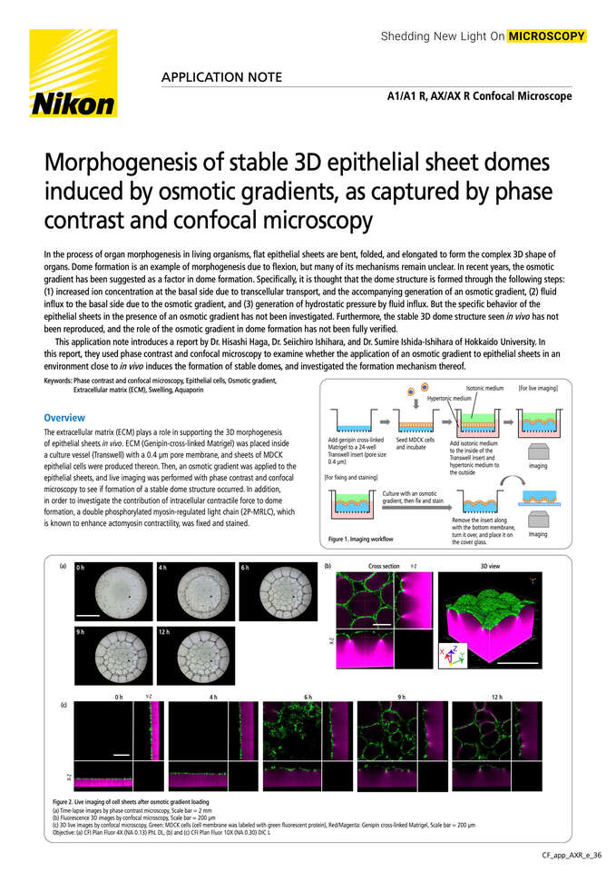
In the process of organ morphogenesis in living organisms, flat epithelial sheets are bent, folded, and elongated to form the complex 3D shape of organs. Dome formation is an example of morphogenesis due to flexion, but many of its mechanisms remain unclear. In recent years, the osmotic gradient has been suggested as a factor in dome formation. Specifically, it is thought that the dome structure is formed through the following steps:
(1) increased ion concentration at the basal side due to transcellular transport, and the accompanying generation of an osmotic gradient, (2) fluid influx to the basal side due to the osmotic gradient, and (3) generation of hydrostatic pressure by fluid influx. But the specific behavior of the epithelial sheets in the presence of an osmotic gradient has not been investigated. Furthermore, the stable 3D dome structure seen in vivo has not been reproduced, and the role of the osmotic gradient in dome formation has not been fully verified.
This application note introduces a report by Dr. Hisashi Haga, Dr. Seiichiro Ishihara, and Dr. Sumire Ishida-Ishihara of Hokkaido University. In this report, they used phase contrast and confocal microscopy to examine whether the application of an osmotic gradient to epithelial sheets in an environment close to in vivo induces the formation of stable domes, and investigated the formation mechanism thereof.