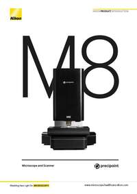
PreciPoint Microscope and Slide Scanners
Products and Promotions may differ based on your selected Region.
Return to the top of the page
Prodotti fuori produzione
Nikon BioImaging Labs provide contract research services for microscope-based imaging and analysis to the biotech, pharma, and larger research communities. Each lab's full-service capabilities include access to cutting-edge microscopy instrumentation and software, but also the services of expert biologists and microscopists, who are available to provide quality cell culture, sample preparation, data acquisition, and data analysis services.
Cliccate qui per iscrivervi alla mailing list di Nikon ed essere i primi a conoscere i nostri nuovi prodotti e le nostre offerte esclusive!
Il Nikon Instruments Learning Center fornisce tutorial interattivi su una varietà di argomenti che vanno da quelli di base a quelli avanzati.
Guide passo passo per l'allineamento e l'utilizzo di microscopi Nikon selezionati.
Trova l'obiettivo Nikon giusto per il tuo flusso di lavoro.
Scarica software e programmi firmware per i prodotti per microscopi Nikon.
Search and filter over 125,000 Open Access Articles that utilize Nikon products and supported third party systems.
MicroscopyU di Nikon è una delle principali fonti di informazioni educative sulla microscopia ottica.
Il sito Web Nikon Small World: tutto sui concorsi di immagine/video al microscopio
Operating modes tailored to diverse workflows
Live-mode, InstantScan mode, SlideScan-mode
Light
Transmitted Light; LED, brightfield
Barcode scanning
Upon request
Supported objectives
20x to 60x air
20x to 100x air and oil
Dimensions
45 cm x 40 cm x 30 cm; 25 kg
Automated Microscope
X-Y-stage automated, Z-axis automated
Handling
Controlled with computer; Live remote control due to automation
Scanning parameters
Whole Slide Imaging or partial digitization
Scanning algorithms
Different scanning algorithms tailored to different slide qualities
InstantScan-mode
Large field of view within seconds at high resolution
Scanning speed per slide
20x: 2 min
100x: 1 hr20x: 2 min
Scanning resolution per slide
60x: 0.85 NA: 0.16 μm/px40x: 0.75 NA: 0.24 μm/px20x: 0.50 NA: 0.479 μm/px
100x: 1.3 NA: 0.096 μm/px60x: 0.85 NA: 0.160 μm/px40x: 0.75 NA: 0.240 μm/px20x: 0.5 NA: 0.479 μm/px
Slide capacity
25 x 75 mm (2 slides) or 50 x 75 mm (1 slide)
Z-Axis Resolution
25 nm
XY-Axis Resolution
78 nm
Z-Stacking
Yes
Operating software
MicroPoint included
Viewer software
ViewPoint* included (unlimited users)
Storage
PreciCloud slide storage
Image analysis
Several software applications on request, based on customer needs
Computers
Various computers recommended and approved by PreciPoint
Microscope computer connection
USB 3.0
Image output formats
PNG, JPEG, TIFF, BMP, VMIC, XLS, and many more
Download 608KB