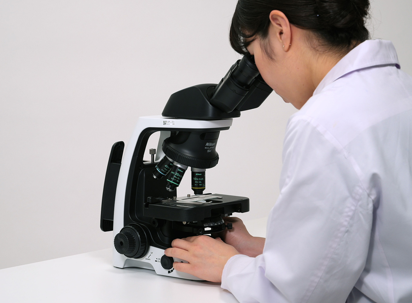
13.ObservationMake observations using phase contrast microscopy
What is phase contrast microscopy?
Phase contrast microscopy is a technique that uses transmitted illumination to observe unstained, colorless and transparent specimens such as living cells.
Insert a phase contrast slider with a diaphragm ring in the slot on the condenser side. It becomes phase contrast microscopy by inserting a phase contrast objective lens with the same PH code into the optical path.
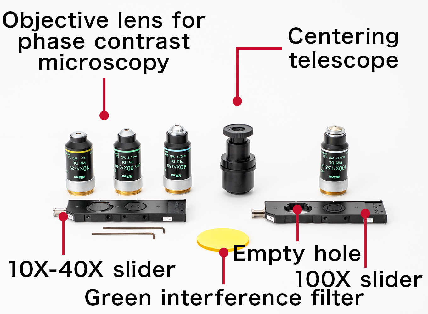
Please ensure the following optical components are available.
10X-40X slider (Microscope slider for phase contrast)
This slider is for 10X, 20X and 40X phase contrast objective lenses. When performing phase contrast microscopy with these objectives, remove the dummy slide from the condenser slider slot and insert this slider.
100X slider (Microscope slider for phase contrast)
This slider is for the 100X phase contrast objective lens. This slider has a empty slot for brightfield observation when using this lens. When switching to phase contrast microscopy, move the slider so the 100x phase ring is in the optical path.
Objective lens for phase contrast microscopy (PH objective lens)
When attaching a phase objective to the nosepiece, check that the PH code of the objective lens matches the PH code of the slider.
Table for the corresponding phase contrast sliders and objective lenses
Phase contrast slider
Phase Objective lens
PH code
Corresponding NA
10X - 40X
PH1
0.25 to 0.5
PH2
0.55 to 8.5
100X
PH3
0.9 to 1.4
Green interference filter
When attached to the field lens during phase contrast microscopy, the contrast of the phase contrast image is improved.
Centering telescope
Used to view the position of the condenser phase ring with the objective phase ring.
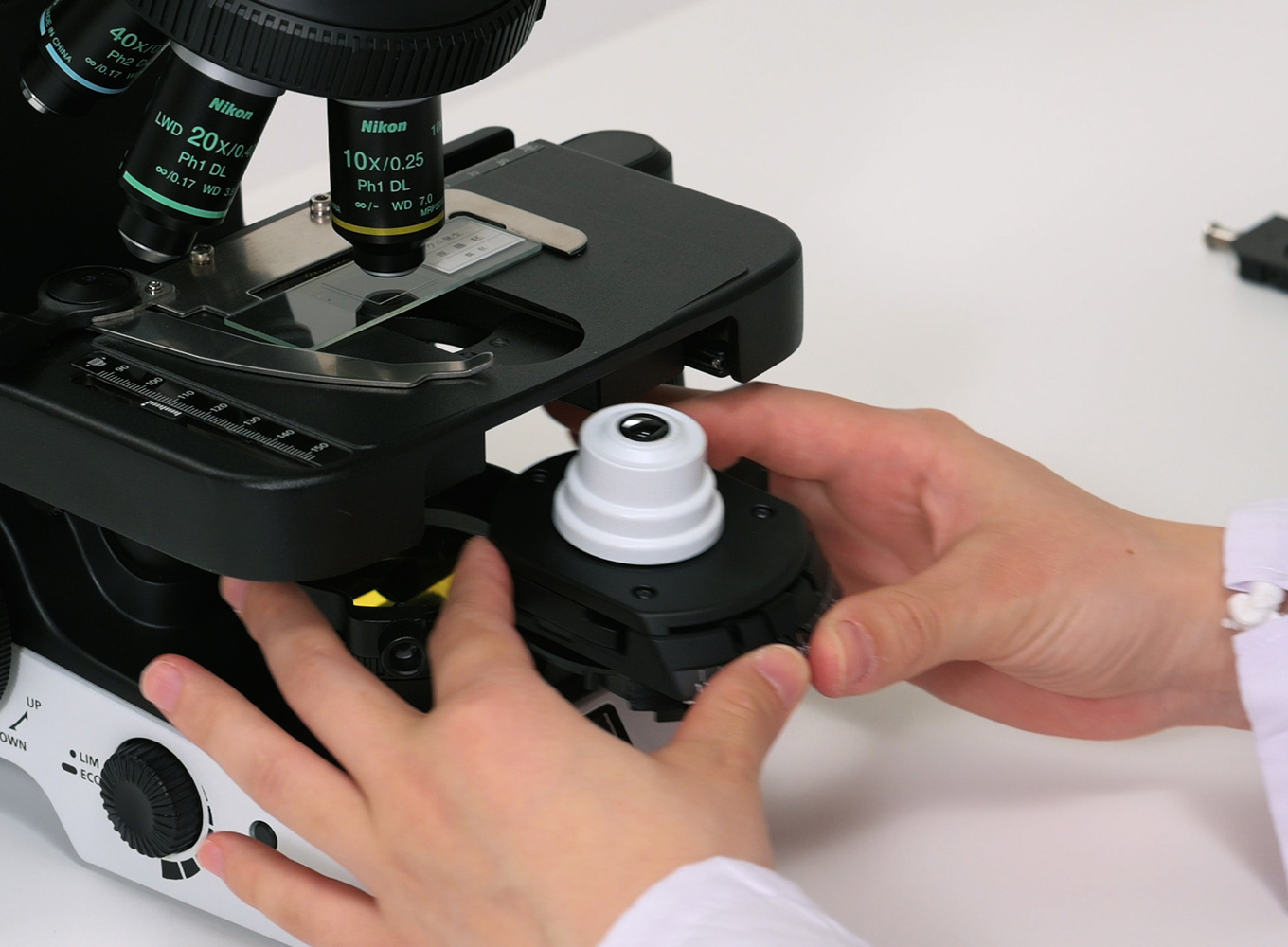
Raise the stage, lower the condenser by adjusting the condenser vertical position knob, then remove the condenser.
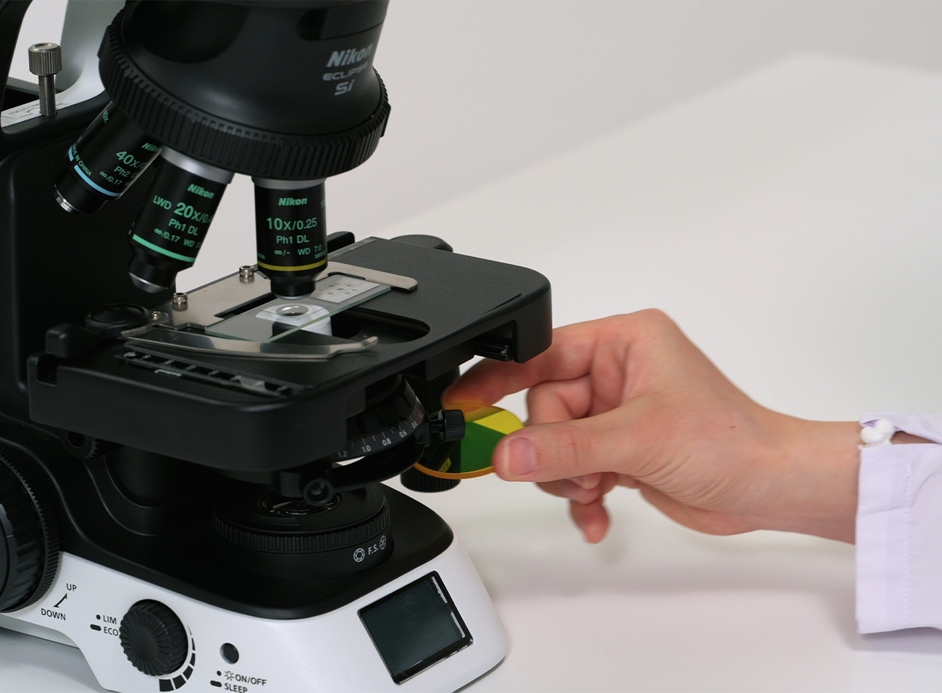
Remove the dummy slider from the removed condenser and insert the 10X-40X phase slider for phase contrast observation.
Return the condenser to its original position and fully open the condenser aperture diaphragm.
Adjust the slider to PH1, then push the slider until you hear a click.
Fully open the field diaphragm and move the condenser vertical position knob to adjust its position until the phase ring is in focus.
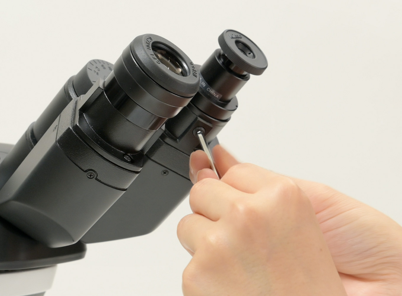
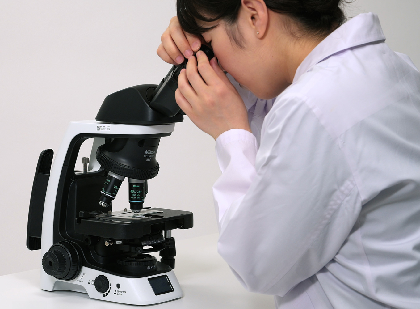
Attach the centering telescope and adjust its focus to view the objective phase ring. Adjust the position of the condenser phase ring to align with the objective phase ring using your dominant eye.
* The eyepiece lens section setting screw is made of resin. Be careful not to tighten it too tightly.
Switch to a 40X objective lens and adjust the slider to PH2, then perform centering again.
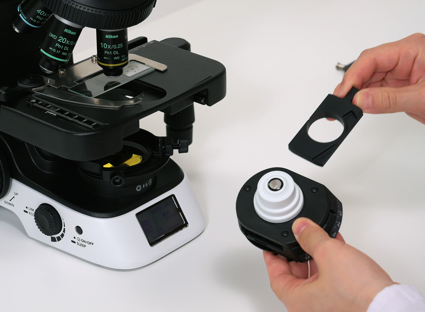
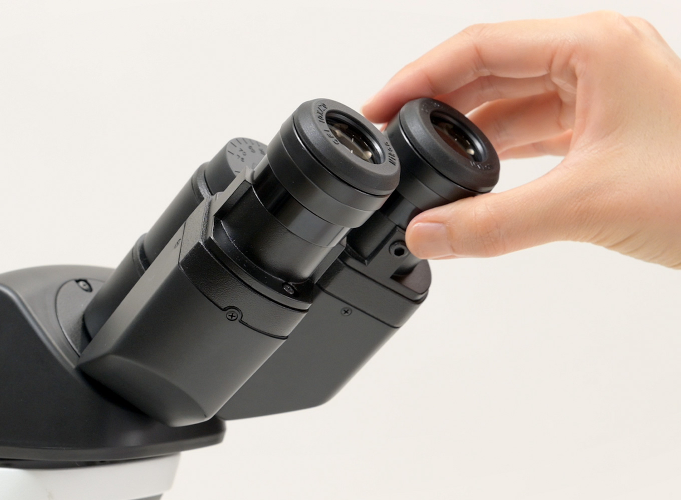
Remove the centering telescope and replace it with an eyepiece lens.
Switch to a 10X objective lens and adjust the slider to PH1.
Place a GIF filter on top of the microscopes field lens.
Focus on the specimen and perform phase contrast observation.
To perform 40x phase contrast observations, rotate that objective into the optical path and move the phase slider to PH2. Refocus the microscope as required.
Microscopy using a phase contrast condenser
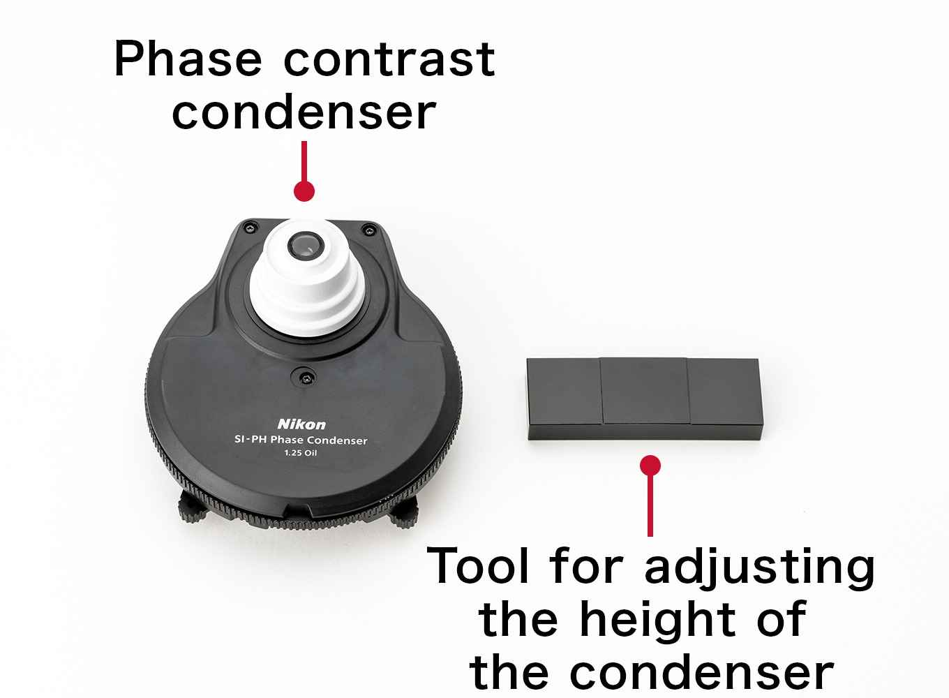
Attachment of the phase contrast condenser and preparation for observation
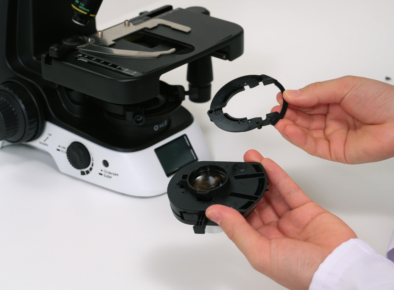
In order to incorporate the Si phase contrast condenser into the ECLIPSE Si, the inner frame component must be removed from the existing built-in condenser and replaced with the Si phase contrast condenser.
Raise the stage and lower the condenser. Loosen the condenser centering knobs on both sides and remove the condenser.
Remove the inner frame component of the condenser attachment section on the rear face of the removed condenser using the hexagonal wrench supplied with the ECLIPSE Si.
Attach the inner frame component of the condenser attachment section using the assembly screws supplied with the Si phase contrast condenser.
Insert the near side of the Si phase contrast condenser into the holder while pressing the bottom of the Si phase contrast condenser against the back of the holder.
At this time, check that the protrusion on the inner frame component of the condenser attachment section fits into the groove of the condenser holder.
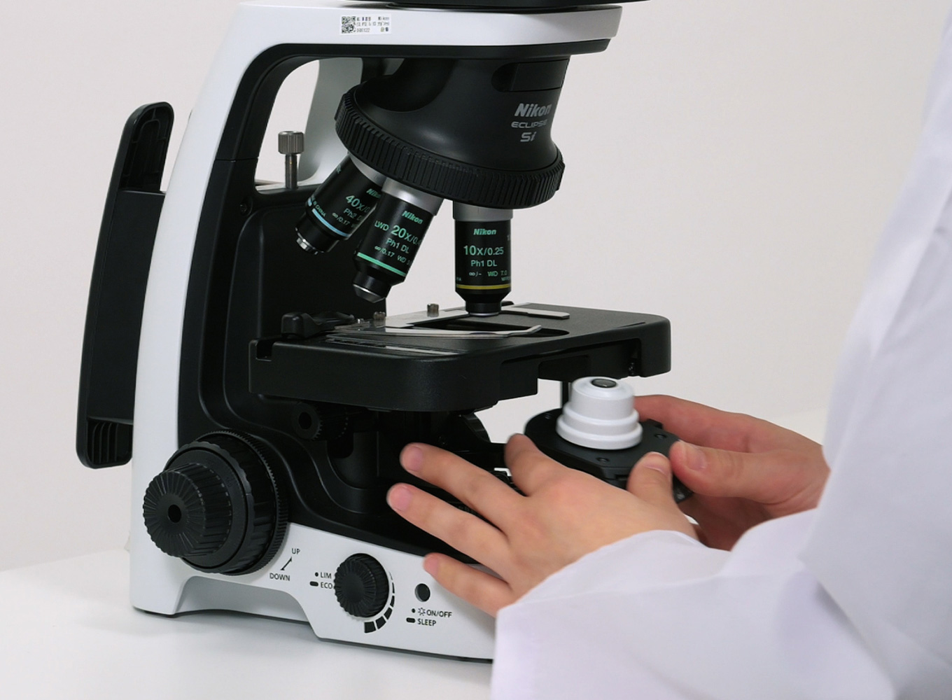
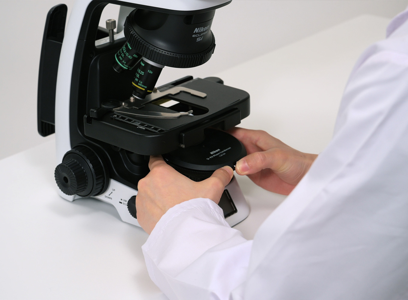
Rotate the stage movement knob to move the stage forward up to the position where the hexagonal height fixing screw can be seen. Loosen the height fixing screw using the supplied hexagonal wrench.
Press the convex part of the height adjustment tool against the opening in the center of the stage and adjust the height of the Si phase contrast condenser using the condenser vertical movement knob so that the bottom surface of the height adjustment tool comes into contact with the upper surface of the condenser lens.
Tighten the hexagonal height fixing lens to fix the height of the condenser.
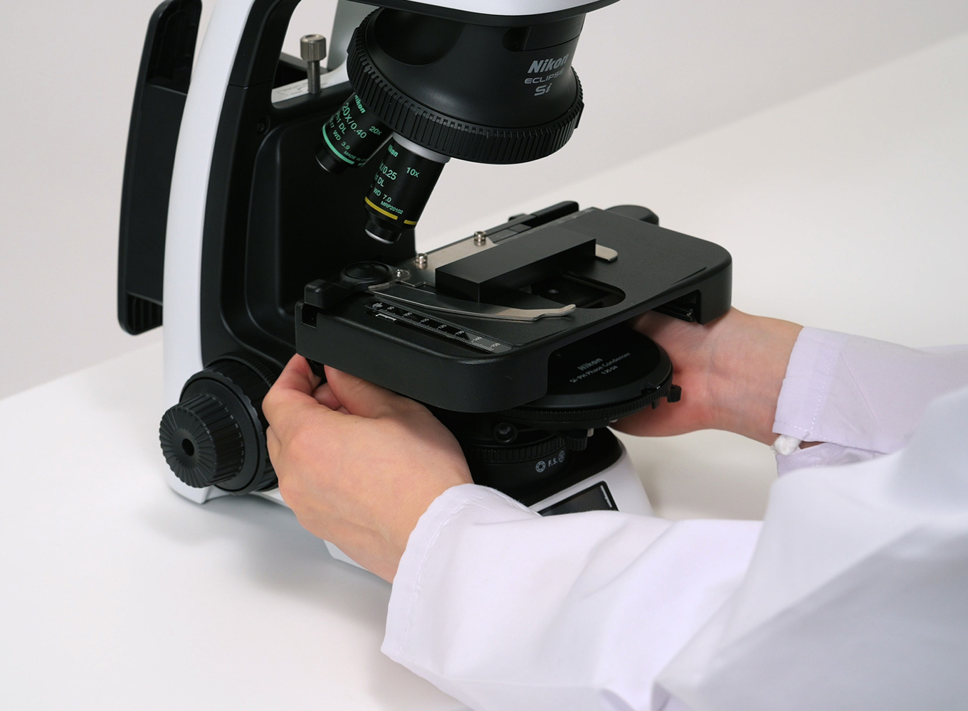
Performing observation using the phase contrast condenser.
Rotate the condenser turret and adjust the position display to [A] (hollow), then adjust the focus using a 10X objective lens for phase contrast observation, while refering to "Preparation For Observation".
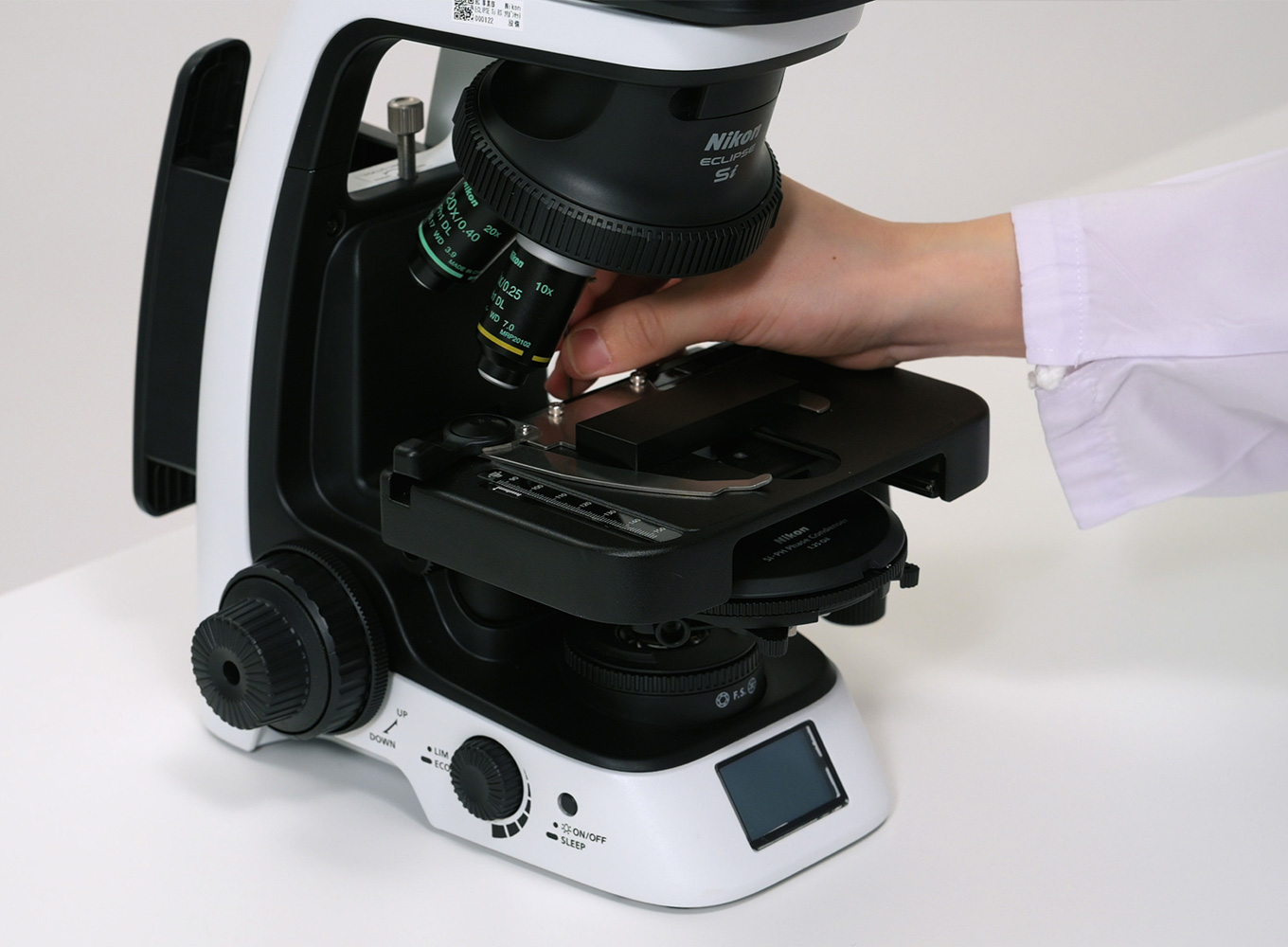
Rotate the condenser turret to adjust it to PH1.
Adjust the field diaphragm dial and condenser vertical movement knob to focus.
Attach the centering telescope and focus on the phase ring of the objective lens, then perform aperture centering using your dominant eye.
*The eyepiece lens setting screw section is made of resin. Be careful not to tighten it too tightly.
Switch to a 40X objective lens and adjust the turret to PH2, then perform centering again.
Remove the centering telescope and attach an eyepiece lens, then switch to a 10X objective lens and adjust the turret to PH1.
Attach a GIF filter to the field lens section.
Focus on the specimen and perform phase contrast observation.
Move the area you want to observe in detail to the center of your field of vision and perform observation by increasing the magnification of the objective lens.