Nikon Europe B.V. | Europe & Africa
- es Change Region
- Global Site
Nikon has established a number of facilities around the world to help local scientific communities gain access to the latest bioscience imaging and optical technologies. These centers and laboratories enable industry professionals, researchers, scholars, and trainees to enhance their research capabilities and provide Nikon with feedback for future product development.
Nikon Imaging Centers and Centers of Excellence are state-of-the-art imaging facilities established as a partnership between key research institutes around the world and Nikon. The mission of these centers is to contribute to the development of cutting edge research and to serve as a hub for educating the surrounding community on the fundamentals of microscopy and the latest advances in imaging technology.
Nikon Microscopy Ambassadors are esteemed scientists, researchers, educators, and professionals who have demonstrated excellence in their respective fields of microscopy. They are passionate advocates for advancing scientific discovery and innovation through the use of Nikon microscopy solutions. They conduct groundbreaking research, educate future generations of scientists, and collaborate with Nikon to develop cutting-edge microscopy technologies.
Nikon Ambassador for Multiphoton & Intravital Imaging. Professor of Intravital Molecular Imaging at the University of Munster. Unparalleled experience and knowledge of imaging challenges in tissue and intravital multiphoton imaging on a wide variety of model organisms validated by numerous high-impact scientific publications.
System portfolio: Nikon's AX R MP multiphoton system
Research areas:
Nikon Ambassador for Cell Imaging and Analysis for Neuroscience. Head of research on the neurobiology of aging at the Centre for Psychiatric Neurosciences and the Leenaards Memory Centre (Lausanne University Hospital - CHUV)
System Portfolio: ECLIPSE Ji with AX Confocal, ECLIPSE Ti2; ECLIPSE Ni-E with Crest-V3, extensive use of GA3 and JOBS
Research Areas:
Nikon Ambassador for Advanced 3D Imaging in Mechanobiology. Research Francqui Professor and Head of the SYMBIOSE Lab at the University of Mons. President of the Research Institute for Biosciences.
System portfolio: Nikon ECLIPSE Ji-AX, Nikon ECLIPSE Ti2E-A1R HD25 confocal microscope, Nikon C1 confocal, NIS-Elements AR with GA3 and JOBS
Research areas:
Website: SYMBIOSE Lab
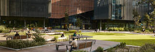
 Nikon BioImaging Lab
 Center of Excellence
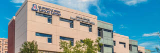
 Nikon Imaging Center
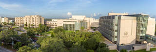