- es Change Region
- Global Site
- Casa
- Nikon BioImaging Centers
- NOLIMITS Unitech

Centro de excelencia
NOLIMITS Unitech
The NOLIMITS Center of Excellence for Plant Biology and Other Life Sciences is the result of a fruitful collaboration between Nikon and the Università degli Studi di Milano. A group of biologists, physicists, and chemists with the support of experienced technicians have developed the microscopy facility within the last 7 years. Such a team of experts now support researchers in different fields of life sciences which primarily include, plant biology, neurosciences, developmental biology and microbiology.
The Center offers microscope solutions allowing the users to investigate in vivo relevant biological processes from single cells, tissues and entire organisms and allowing super-resolution analyses. The multidisciplinary scientific interests of the users of the center guarantee a continuous exchange of ideas in which the technical staff acts as a hub by putting the different realities in communication with each other. The large audience of plant biologists that have access to the center and their expertise has led to the choice of performing customization of the microscopes with solutions dedicated to plant specimens (e.g. water and silicon immersion objectives), but perfect also for other large specimens.
The Nikon Center of Excellence at UNIMI brings these cutting-edge techniques to all researchers, in Milan and beyond, with expert advice on how to choose the best technique for a given question and project. Specific development of new sample preparation, imaging and image analysis and processing techniques is performed based on the Center of Excellence microscopes within the facility. The use of microscopy coupled with the use of organisms expressing genetically encoded biosensors is another prerogative of this Nikon Center of Excellence.
Contacto
Academic Director
email hidden; JavaScript is required
Facility Manager
email hidden; JavaScript is required
Address
Università degli Studi di MilanoVia C. Golgi 19 - 20133 Milano, Italy
Website
Systems Available
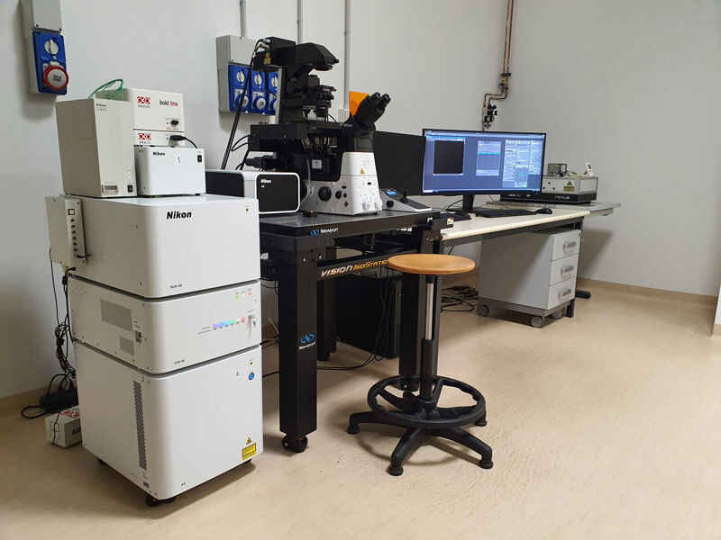
Nikon ECLIPSE Ti2-E with AX R Confocal
The application fields of the Nikon AX R 2K HD laser scanning confocal inverted microscope range from cellular biology to histology, from physiology to material sciences. The presence of a high-speed resonant scanner allows the observation of cellular kinetics with an excellent temporal resolution. Also, the fast resonant scanner can be used for live and gentle imaging by minimizing phototoxicity and photobleaching.
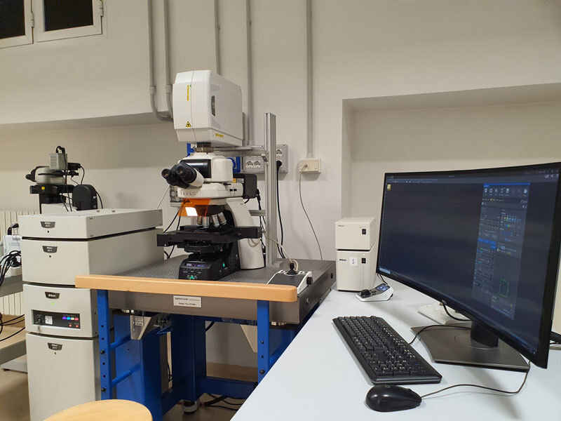
ECLIPSE Ni-E with A1 Confocal and PicoQuant FLIM
The ECLIPSE Ni-E A1 laser scanning confocal upright microscope is used in all fields of application of biological and microbiological interest, but also in chemistry and in the material sciences. The DUVB emission spectral detector allows the definition of collecting windows for fluorescence signals based on the user’s experimental needs and also allows you to perform spectral acquisition and separation experiments. The PicoQuant module allows you to perform FRET-FLIM and FCS experiments with two highly sensitive Hybrid Detectors.
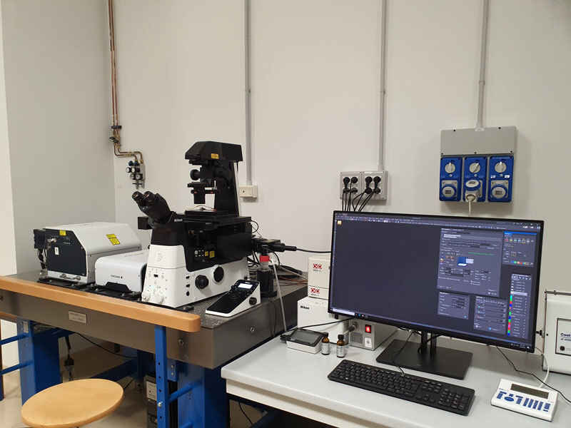
ECLIPSE Ti2-E with Yokogawa W1-SoRa
The Yokogawa spinning disk (CSU-W1-SoRa) is equipped with 405, 488, 561, and 647 nm laser lines and a multiband filter for fast-triggered acquisitions of up to 4 channels. The spinning disk detector is a Photometrics BSI sCMOS (6.5 μm pixel with 93% QE) camera, allowing for fast, high-resolution imaging. The SoRa module increases resolution by 1.4X optically and with deconvolution up to 2X improvement compared to standard confocal microscopes.
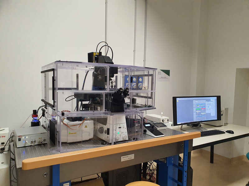
ECLIPSE Ti2-E Inverted Microscope with N-SIM E Super-Resolution
The Nikon ECLIPSE Ti2-E inverted microscope is used in all fields of application of biological and microbiological interest, but also in chemistry and in the material sciences. In many cases with the same sample, you can easily perform acquisitions in the three different modes allowed by the instruments: widefield, confocal and super-resolution in a fast and intuitive way. The N-SIM E super-resolution module allows for reaching a lateral resolution of 115 nm and an axial resolution of 269 nm.
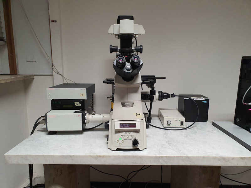
ECLIPSE Ti-E Widefield Inverted Microscope
The ECLIPSE Ti-E is a system for high-speed widefield microscopy ideal for live cell imaging. The CoolLED pE800 includes 8 LED sources which allow for flexible fluorescence imaging. The system is equipped with a Hamamatsu ORCA D2 (dual sensor camera) suitable for FRET analyses (CFP/YFP and GFP/RFP). The Okolab incubator system for temperature, CO2 and humidity control allows the setting up of in vivo experiments.
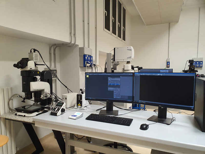
SMZ18 Stereo Microscope
In various fields such as embryology & developmental biology, there is a growing need for imaging systems that range at different scales: from single cells to entire organisms. The SMZ18 was created as a response to this need and is also used in the fields of material sciences, mineralogy, archaeology and in the study of cultural heritage. The stereo is equipped with the CoolLED pE-300ultra and two cameras (DS-Fi3 color and Hamamatsu Orca LT3) for both brightfield and fluorescence observations.
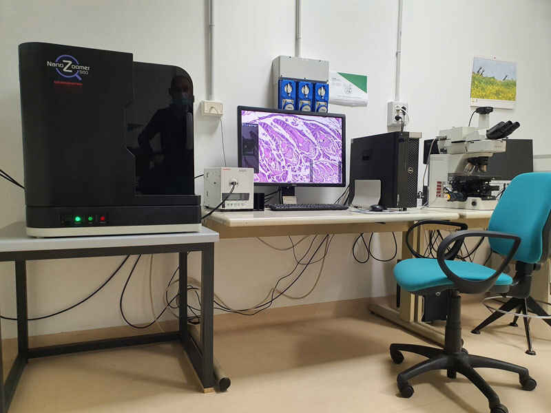
Hamamatsu NanoZoomer S60 Slide Scanner
The Hamamatsu NanoZoomer S60 slide scanner belongs to the family of digital slide scanners that convert slides into high-resolution digital images by high-speed scanning in both brightfield and fluorescence. Being a versatile instrument, the NanoZoomer S60 is widely employed in different fields, from basic to applied research, proving to be valuable also in pathology and clinical diagnosis fields.
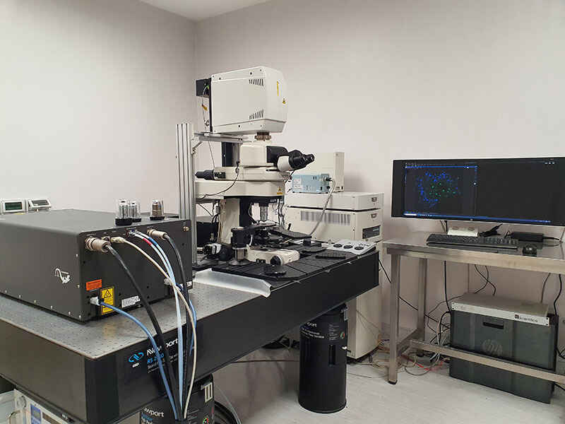
ECLIPSE Ni with A1 MP+ Multi and Single-Photon
The A1 MP+ multiphoton microscope enables high-speed, deep-tissue imaging in live and fixed organisms with unprecedented sensitivity and clarity. The microscope base is an upright Nikon ECLIPSE Ni, which allows repeatable focusing in the Z-axis when large movements are required. The multiphoton laser is a Coherent dual IR pulsed laser (820-1300 nm + 1040 nm). The scanning head has 4 GaAsP detectors + 1 DUVB spectral GaAsP detector and resonant 1K HD scanner.
- Casa
- Nikon BioImaging Centers
- NOLIMITS Unitech
