Products and Promotions may differ based on your selected Region.
Return to the top of the page
Produtos descontinuados
Nikon BioImaging Labs provide contract research services for microscope-based imaging and analysis to the biotech, pharma, and larger research communities. Each lab's full-service capabilities include access to cutting-edge microscopy instrumentation and software, but also the services of expert biologists and microscopists, who are available to provide quality cell culture, sample preparation, data acquisition, and data analysis services.
Search and filter over 125,000 Open Access Articles that utilize Nikon products and supported third party systems.
O Denoise.ai remove automaticamente o ruído de Poisson das imagens confocais - experimente gratuitamente.
Baixe programas de software e firmware dos produtos para microscópio da Nikon.
Site da Nikon Small World - tudo sobre as competições de imagens/vídeos adquiridos por um microscópio.
Guias passo a passo para alinhar e operar os microscópios Nikon selecionados.
MicroscopyU da Nikon é uma fonte importante de informações educacionais sobre microscopia óptica.
Encontre a objetiva Nikon certa para o seu fluxo de trabalho.
agosto 2023
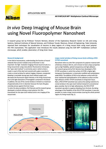
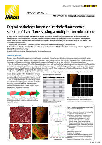
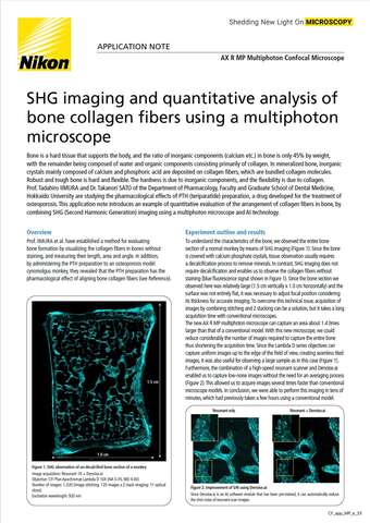
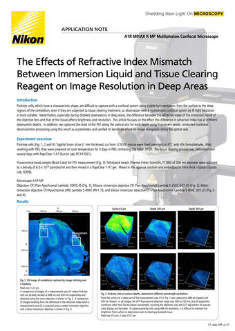
Leia mais e baixe
abril 2023
dezembro 2022
outubro 2022
junho 2022
abril 2022
agosto 2021
outubro 2020
dezembro 2015
Baixar 11.19MB