Nikon Corporation Healthcare Business Unit | Asia, Oceania & Middle East
- en Change Region
- Global Site
September 2025
The intestinal tract is one of the most important absorptive organs in the body, and its luminal surface exhibits a characteristic three-dimensional structure composed of crypts and villi. Intestinal epithelial stem cells located at the base of the crypts differentiate into absorptive epithelial cells, goblet cells, and Paneth cells, thereby supporting the diverse physiological functions of the intestine. In particular, goblet cells secrete mucin to form a protective mucus layer. This intestinal-mucosal surface plays a crucial role in the maintenance of intestinal homeostasis and symbiosis with gut microbiota.
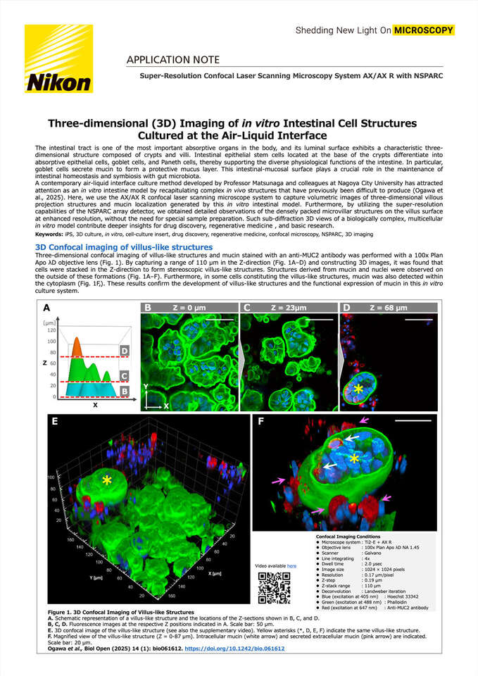
A contemporary air-liquid interface culture method developed by Professor Matsunaga and colleagues at Nagoya City University has attracted attention as an in vitro intestine model by recapitulating complex in vivo structures that have previously been difficult to produce (Ogawa et al., 2025). Here, we use the AX/AX R confocal laser scanning microscope system to capture volumetric images of three-dimensional villous projection structures and mucin localization generated by this in vitro intestinal model. Furthermore, by utilizing the super-resolution capabilities of the NSPARC array detector, we obtained detailed observations of the densely packed microvillar structures on the villus surface at enhanced resolution, without the need for special sample preparation. Such sub-diffraction 3D views of a biologically complex, multicellular in vitro model contribute deeper insights for drug discovery, regenerative medicine , and basic research.
Keywords: iPS, 3D culture, in vitro, cell-culture insert, drug discovery, regenerative medicine, confocal microscopy, NSPARC, 3D imaging