
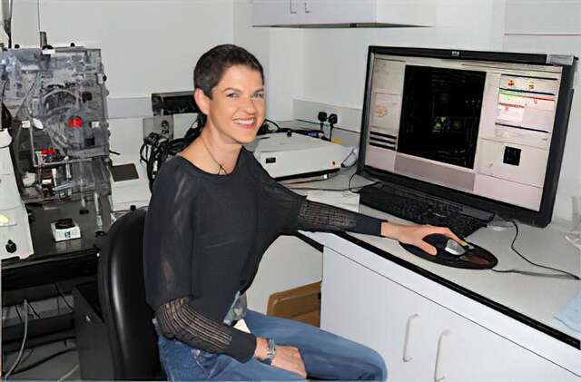
Smart Automation with Confocal Microscope: ECLIPSE Ji Changes the Future of 3D Cell Research
AdHuCell platform and Research Institute for Biosciences
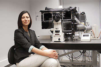
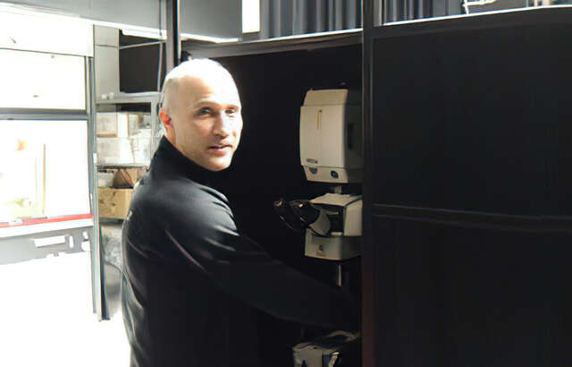
University of Mons - UMONS
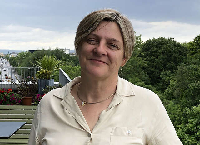
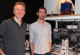
Professor Sylvain Gabriele, SYMBIOSE Lab
Professor Sylvain Gabriele at the University of Mons (UMONS), Belgium, is a leading researcher in mechanobiology, investigating how cells sense, respond to, and retain memory of mechanical forces.
In this interview, we spoke with Professor Gabriel about how the Nikon ECLIPSE Ji-AX Confocal Integrated System has advanced their understanding of complex 3D biological systems.

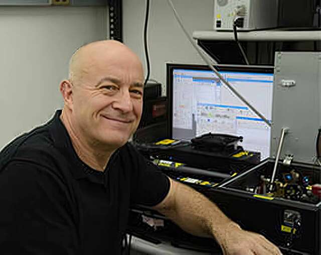
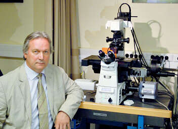
持久卓越 - 经久耐用的显微技术如何推动LIV 的创新
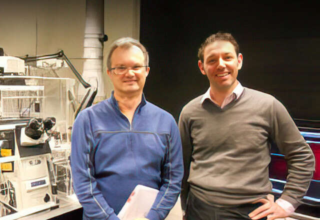
Christian Conze博士
汉堡莱布尼茨病毒学研究所(LIV)光学显微镜与图像分析技术平台负责人
为庆祝尼康显微仪器诞生100周年,我们推出了“持久卓越”(Enduring Excellence)系列活动,旨在向那些长期使用尼康系统的研究人员致敬,他们的使用经验充分彰显了尼康设备所蕴含的可持续性与持久价值。
在本期内容中,我们把目光投向了汉堡莱布尼茨病毒学研究所(LIV)尼康卓越中心的光学显微镜与图像分析技术平台负责人Christian Conze,在他的整个科研历程中,他始终信赖并使用着经久耐用的尼康显微镜。

历经时间考验的性能 - VIB-KU Leuven的实践案例
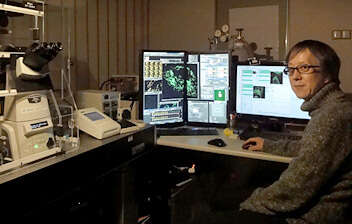
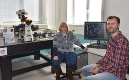
尼基·科尔索特(Nikky Corthout)
光学显微镜专家 - VIB生物成像核心实验室鲁汶,VIB-KU鲁汶脑与疾病中心
帕布罗·埃尔南德斯·巴拉斯(Pablo Hernández Varas)
实验室负责人 - VIB生物成像核心实验室鲁汶,VIB-KU鲁汶脑与疾病中心
一个多世纪以来,尼康显微镜系统一直以持久耐用为设计理念。在本期我们采访了来自比利时鲁汶大学尼康卓越中心的VIB生物成像核心实验室的帕布罗·埃尔南德斯·巴拉斯和尼基·科尔索特。两位学者一直在使用实验室配备了17年以上的尼康A1 R共聚焦显微镜系统。他们的见解不仅突显了尼康显微系统的耐用性,还展示了该系统如何持续支持前沿的科学发现。


客户访谈 - ECLIPSE Si助力提升眼科医疗水平
“外形小巧、操作简单、画质优良即是选择的理由”
尼康生物显微镜ECLIPSE Si,基于人体工程学,旨在实现自然姿势下的观察和提高操作效率。爱媛县“石鎚眼科”针对结膜炎等眼疾的治疗,引入“眼脂涂抹检查”法,并在检查中使用了ECLIPSE Si。我们就选用该显微镜的原因以及实际操作感受等,采访了“石鎚眼科”理事长铃木崇(Suzuki Takashi)医生。
※本产品在日本国内并不属于医疗器械。同时,本页所刊登的就本产品对医生进行采访的内容,并非用以保证本产品的效能、效果及性能,也并不表示该医生公开认可、推荐、指导或选用本产品。

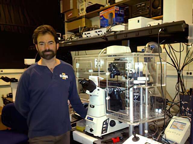
YOUR SWITCH TO THE FUTURE
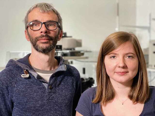
巴塞尔大学生物医学系 (DBM)
Pascal Lorentz
Michael Abanto博士
尼康卓越中心主任
显微镜核心器件负责人
研究大纲:免疫学和传染病、神经科学和癌症生物学以及组织发育和再生

Dr. Kevin Richetin
尼康显微镜形象大使Kevin Richetin博士是一位研究阿尔茨海默氏症和开发神经退行性疾病工具的神经学家。在这段采访视频中,Dr. Richetin 谈到,他们之所以选择尼康显微镜,是因为其软件NIS-Elements 可以实现分析和自动化的无缝集成。他强调,这种独特的功能对于长时间以极高分辨率跟踪颗粒而言极为有用,这也是尼康的与众不同之处。

研究人员访谈 - YOUR SWITCH TO THE FUTURE
辛辛那提儿童医院医疗中心
加州大学儿科教授
Matthew Kofron 博士
辛辛那提囊胞性纤维症治疗研究中心
Alicia Ostmann
武部研究室 肠胃病学部
岩泽坚太郎
研究大纲:改善儿童健康。制备类器官。评估囊胞性纤维症患者用药的疗效。

研究人员访谈 - YOUR SWITCH TO THE FUTURE
European Institute for Molecular Imaging (EIMI)
体内分子成像部
Friedemann Kiefer 教授(博士)
研究大纲:阐明分子机制如何在生物学上塑造人体细胞和组织。多尺度成像。


Nikon BioImaging Lab contributes to “mini-gut” research
Dr. Hidenori Akutsu
Director of the Department of Reproductive Medicine, Center for Regenerative Medicine, National Center for Child Health and Development
Nikon Equipment and Services

YOUR SWITCH TO THE FUTURE
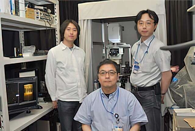
大阪大学 微生物病研究所
环境应答研究部门 细胞控制领域
三木裕明 教授
船户洋佑 副教授
桥爪脩 助理教授
研究大纲:分析称为细胞周期蛋白 M 功能,该分子在从细胞中排出镁离子方面发挥作用。

The ECLIPSE Ci-L plus contributes to more comfortable clinical examinations.
“Together with the high optical performance, I realized its thoughtful design aids our daily work.”
Clinical examinations play an important role in medical care. Nikon has developed a new biological microscope, the ECLIPSE Ci-L plus, with the concept of reducing physical and mental strain on clinicians and laboratory technicians who use microscopes on a daily basis. In this interview, we talked to Dr. Akira Yoshikawa from the Department of Anatomic Pathology, Kameda Medical Center – a flagship hospital in the southern part of Chiba prefecture, Japan – on his thoughts after employing this microscope and the newly developed objective lens for microscopes ‘CFI Plan Apochromat Lambda D’ for practical everyday use.
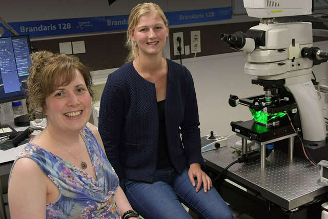
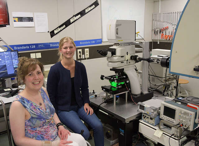
Associate Prof. Dr. Klazina Kooiman and Dr. Ines Beekers
Associate Prof. Dr. Klazina Kooiman, Head of Therapeutic Ultrasound Contrast Agent Group, and Dr. Ines Beekers, Postdoctoral Researcher in the Department of Biomedical Engineering of the Thoraxcenter, Erasmus MC, Rotterdam, the Netherlands
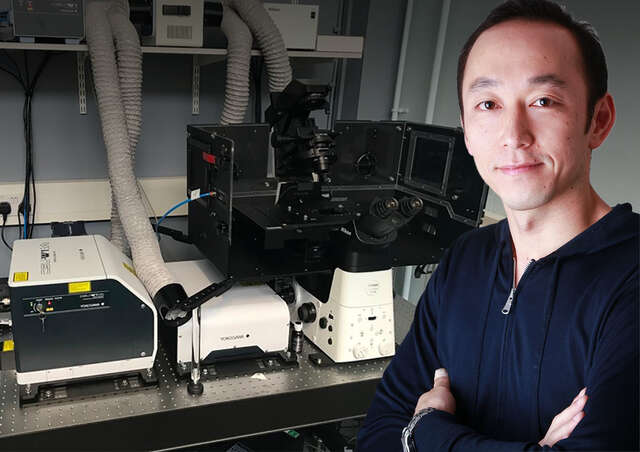
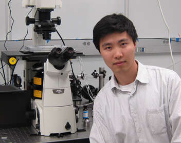
Dr. Yohei Yamauchi
Dr. Yohei Yamauchi, Principal Investigator, Cell biologist of viral infections, School of Cellular and Molecular Medicine, University of Bristol, UK
Assistant Prof. Joseph Michael Hyser & Dr. Alexandra Leigh Chang-Graham
Virology and Microbiology
Baylor College of Medicine
Houston, Texas, USA
Assistant Prof. Klazina Kooiman & Inés Beekers
Therapeutic Ultrasound Contrast Agent Group, Thoraxcenter, Department of Biomedical Engineering, Erasmus MC, Rotterdam
Dr. Steven Nedellec and Dr. Tiphaine Douanne
Dr. Steven Nedellec, Facility Manager of MicroPICell, Université de Nantes, France and Dr. Tiphaine Douanne, Universite de Nantes, Signaling in Oncogenesis, Angiogenesis and Permeability, CRCINA INSERM U1232, France
Ronald D. Vale, Ph.D.
Professor and Vice-Chairman of the Department of Cellular and Molecular Pharmacology
Investigator, Howard Hughes Medical Institute (HHMI)
The University of California, San Francisco San Francisco, CA, USA
Tamas Freund, Ph.D. and Istvan Katona, Ph.D.
Institute of Experimental Medicine of the Hungarian Academy of Sciences (IEM HAS)
Budapest, Hungary
Paul Ronald Selvin, Ph.D.
Professor, Department of Physics, University of Illinois at Urbana-Champaign
Atsushi Miyawaki, M.D., Ph.D.
Senior Team Leader, Laboratory for Cell Function Dynamics, RIKEN Brain Science Institute
Ikuo Wada, Ph.D.
Department of Cell Science, Institute of Biomedical Science, Fukushima Medical University
Romain Le Bars,博士
Imagerie-Gif光学显微核心机构设施是法国生物成像基础设施的一部分。该机构由法国Gif sur Yvette 细胞综合生物学研究所 (I2BC)主持。
StaffanStrömblad教授,博士
StaffanStrömblad博士是著名的瑞典斯德哥尔摩卡罗林斯卡学院(Karolinska Institutet)的主管,年度诺贝尔生理学奖正是由该学院颁发。他还是Live Cell Imaging Facility(LCI)的负责人,该机构整合了尼康活细胞成像中心。
Dr. Arne Seitz, Dr. Romain Guiet and Thierry Laroche
The Faculty of Life Science (SV) at the École Polytechnique Fédéral de Laussane (EPFL), Switzerland, has a long record of excellence in research applied to life sciences
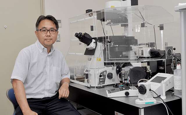
Masato Nakagawa
所谓的iPS细胞作为多能干细胞引起了相当大的研究兴趣,因为它或将在医学界开辟一片全新的天地。京都大学iPS细胞研究与应用中心(CiRA)正在开展广泛的研究活动,旨在利用iPS细胞实现再生医学。这项iPS细胞研究中采用了尼康BioStation CT细胞培养观察系统,为提高研究效率助力。
我们很高兴有机会与在CiRA从事iPS细胞研究的Masato Nakagawa先生进行交流。
注:附列于各研究者侧的机构与职务信息仅代表访谈当时其所在的机构及担任的职务。