Nikon Europe B.V. | Europe & Africa
- pt Change Region
- Global Site
For 100 years, we've explored the microscopic world,
driven by a desire to see and understand.
Our optical technologies help illuminate the nanoscale,
expanding possibilities in science,
medical care, and all that surrounds us.
Lighting a path to a brighter future,
these innovations open the door to new discoveries,
enhancing lives every day.
With the power of light,
we continue to unlock the future.
JOICO 100th Anniversary - NIKON JOICO AWARD Exhibition
Exhibition
From Apr. 1, 2025
Nikon Solutions Co., Ltd. Tokyo, Japan Showroom (Tokyo)
Magnified
Exhibition
From Mar. 15 to Jan. 30, 2026
ARTIS-Micropia, Netherlands
Exhibition of Nikon Small World
Exhibition
From Apr. 19, 2025 to May 3, 2026
Seoul Science Center, South Korea (Seoul, South Korea)
Nikon Small World at the California Science Center
Exhibition
From May 1, 2023 to June 30, 2027
California Science Center (Los Angeles, California)
Special Program for the JOICO 100th Anniversary – "Where Are the Awardees Now?"
Webinar
July 4, 2025 (Fri)
Professor Masatsugu Toyota (Saitama University)
Commemorative Lecture for the JOICO 100th Anniversary
Webinar
July 10, 2025 (Thu)
Shigeo Okabe M.D., Ph.D. (University of Tokyo)
Nobutaka Hattori M.D., Ph.D. (Juntendo University)
Special Program for the JOICO 100th Anniversary – "Where Are the Awardees Now?"
Webinar
August 22, 2025 (Fri)
Tomoe Ishikawa Ph.D. (Massachusetts Institute of Technology)
Special Program for the JOICO 100th Anniversary – "Where Are the Awardees Now?"
Webinar
October 8, 2025 (Wed)
Tetsuo Hasegawa M.D., Ph.D. (University of Cambridge)
JOICO 100th Anniversary - NIKON JOICO AWARD Exhibition
JOICO 100th Anniversary - NIKON JOICO AWARD Exhibition
From Apr. 1, 2025
Nikon Solutions Co., Ltd. Tokyo Showroom (Tokyo, Japan)
Celebrating 100 Years of Microscope Business. Featuring award-winning works and a commemorative message board for visitor comments.
Magnified
Magnified
From Mar. 15 to Jan. 30, 2026
ARTIS-Micropia (Amsterdam, Netherlands)
Exhibition of historical microscopes and Nikon Small World award-winning works
Exhibition of Nikon Small World
Exhibition of Nikon Small World
From Apr. 19, 2025 to May 3, 2026
Seoul Science Center, South Korea (Seoul, South Korea)
Exhibition of Nikon Small World 2024 Award-winning Works
Nikon Small World at the California Science Center
Nikon Small World at the California Science Center
From May 1, 2023 to June 30, 2027
California Science Center (Los Angeles, California)
Exhibition of Nikon Small World and Small World in Motion Annual Award-Winning Collection
We are fascinated by lymphatic vessels, the 2nd vascular system of vertebrates, and try to understand their tissue-specific formation and function as part of the immune system.
We believe that a better understanding of developmental and disease processes in life science requires to study cells and tissues across scales to maintain the context of the entire organ or even organism.
Different microscopes are essential tools in our research. Many of our discoveries of lymph vessel development and malfunction were sparked by discrepancies between published models and our microscopic observations.
To image cells, structures and processes within their tissue context has always been our vision. Development of new labels, transgenic models and microscopes now allows us imaging of whole organs or even in the living organism.
YOUR SWITCH TO THE FUTURE (interview): European Institute for Molecular Imaging (EIMI)
…Alberto Diaspro is a full professor of applied physics, and his research is related to the design, realisation and utilisation of advanced optical microscopy applied to molecular oncology, neuroscience and smart drug delivery.
Label-free and fluorescence optical microscopes are the key elements to answer fundamental questions in biology. 4D (x, y, z, t) imaging of living systems at molecular level is boosted by quantum based sources and detectors with AI.
Thanks to my achievements in this field, I received the 2022 Weber prize for excellence in fluorescence and the 2024 Fermi prize for notable original contributions to the development and application of optical microscopy and the crucial impact on cellular and molecular biophysics.
…My work strongly focuses on enabling researchers to make educated decisions when designing their microscopy experiments. We actively support them in that endeavor, before, during and after training them to use our equipment.
We aim at enhancing science reproducibility through education. Our pedagogy-based training helps users develop technical understanding and critical thinking. We also educate trainers through Train-the-trainer shadowing programs and courses.
…In my training and in the consolidation of my research lines, fluorescence microscopy has been an essential tool. Without it, I would not have been able to build a scientific career, nor would I have had the opportunity to travel the world and meet passionate researchers and scientists.
New technologies have allowed me to move from in vitro to in vivo studies, made possible above all by advances in imaging techniques. In my research, I shifted from studying calcium dynamics at the single-cell level in isolated tissues to investigating calcium dynamics in single organelles in intact, pot-grown plants. For me, this achievement is truly remarkable.
My goal is to study how plants respond to environmental stimuli in a non-invasive way, that is, under their natural growth conditions. To achieve this, I aim to develop plants expressing biosensors for specific analytes, combined with the new tailored imaging solutions.
…We are developing innovative biophotonic measurement technologies, including optical microscopes that integrate photonic and information technologies. Our focus is on creating techniques capable of acquiring large-scale, high-speed data from biological samples.
The optical microscope itself is at the heart of our research. By examining the fundamental principles behind the imaging process, we are designing new ideas and technologies to extract even more information from living systems.
Free from commercial constraints, we are constantly exploring the ultimate possibilities of optical microscopy as a measurement technology, striving each day to push the boundaries of what can be observed and understood from life itself.
…"Immune cells move around to do their work within the human body. In order to analyze the actual movements of these cells, I am conducting research using multiphoton microscopes to observe living cells in living tissues and living individuals.
No microscopy, no life.
My life as a researcher would not be possible without microscopes. I've always loved cameras, and I never get bored even if I spend half a day observing through a microscope. New visualization techniques are leading to new discoveries in biology. As Nikon's microscopes continue to evolve, I will continue to explore new concepts regarding the dynamic control of immune cells."
"I lead the NeuroCyto lab where we apply advanced microscopy techniques to directly observe nanoscale molecular assemblies in neurons, to understand the physiology of these unique cells.
Smaller, faster, gentler: I aim to push microscopy on all fronts to power new biological discoveries. But in the end, it’s all about working with great people and training the next generation.
As Yogi Berra said, “You can observe a lot by just watching”. Everything I have done and discovered as a scientist has been through the eyepieces (or cameras) of microscopes.
With the advent of super-resolution microscopy, with the breakthrough of AI applications, we can do things today with microscopes that I thought would be impossible when I started almost 25 years ago."
“We apply advanced imaging approaches to study 3D models and tissues to visualise how cells sense and respond to their environment in diseases such as cancer and fibrosis. Microscopes are critical for all our research. They enable exquisite insight into spatial dynamics, interactions and mechanisms across biological scales. We collaborate with excellent imaging teams across the world – a great community to be part of!”
…“Through the establishment of GCEA (Global CHA Embryolab Academy), we have introduced the latest systems and a variety of high-level training programs, laying the foundation for systematic training of embryology researchers. We already use various Nikon microscopes, and in particular, the ECLIPSE Ti2-I has greatly shortened the workflow for micromanipulation. Its various automated functions significantly reduced work fatigue and greatly improved work efficiency, which was very impressive.
Going forward, GCEA will continue to actively adopt such cutting-edge technologies and strive to continually innovate assisted reproductive technology (ART).”
The ECLIPSE Ti2-I supports embryologists training at South Korea’s ART Academy
…“For the past 15 years my research activities have been focused on understanding the disease mechanisms affecting the brain by providing know-how and access to state-of-the-art microscopes.
Microscopes are key to my work. By visualizing targets in the brain by basic slide scanning up to advanced confocal, we answer questions that impact the life of people with neurodegenerative diseases. Some images I use to highlight the beauty of biology and present something new that engages the community.”
Webinar “How to Image and Quantify Human Astrocytes in Alzheimer’s Disease Chimeras with AI”
…“Director of the CCHMC Bio-Imaging and Analysis (BAF). The goal of the BAF is to provide users with cost-effective access to and training on high-end confocal, two-photon, wide-field microscopes, light sheet microscopes and image analysis.
As a director of a shared facility in a large pediatric medical research center, I endeavor to enable investigators to perform research which will ultimately improve child health.”
YOUR SWITCH TO THE FUTURE (interview): Cincinnati Children’s Hospital Medical Center
…“I lead Moffitt’s BioEngineering Department, pioneering 3D tumor modeling, immunotherapy, and bioimaging to accelerate cancer research, improve drug delivery, and develop innovative cancer therapies.
At Moffitt, we envision a future where advanced microscopy and bioengineering come together to create precise, patient-specific 3D tumor models. These models will enable real-time, high-resolution evaluation towards the discovery of personalized cancer therapies.”
…“Our laboratory develops cutting-edge bioimaging technologies and utilizes advanced Nikon microscopes to investigate cellular signaling and functions in cancer cells, neurons, and stem cells. We are also pioneering optogenetics—innovative techniques that precisely control cell functions using light.
By integrating bioimaging, molecular optogenetics, protein engineering, and mRNA regulation technologies, our lab aims to revolutionize fundamental brain science research and propose new paradigms for the treatment of neurological disorders.”
YOUR SWITCH TO THE FUTURE (interview): Korea Advanced Institute of Science and Technology
…“Our lab researches plasmonic nanostructures for sensing, light modulation, and signal enhancement, aiming to develop sensitive, low-cost diagnostic devices with novel functionalities.
We use Nikon Ti2-U and Ti2-E + AX R optical microscopes to build custom platforms for characterizing plasmonic devices and studying light-matter interactions, key to developing ultra-sensitive technologies.”
…University of Münster,
European Institute for Molecular Imaging (EIMI),
Multiscale Imaging Centre, Münster, Germany
We are fascinated by lymphatic vessels, the 2nd vascular system of vertebrates, and try to understand their tissue-specific formation and function as part of the immune system.
We believe that a better understanding of developmental and disease processes in life science requires to study cells and tissues across scales to maintain the context of the entire organ or even organism.
Different microscopes are essential tools in our research. Many of our discoveries of lymph vessel development and malfunction were sparked by discrepancies between published models and our microscopic observations.
To image cells, structures and processes within their tissue context has always been our vision. Development of new labels, transgenic models and microscopes now allows us imaging of whole organs or even in the living organism.
YOUR SWITCH TO THE FUTURE (interview): European Institute for Molecular Imaging (EIMI)
University of Genoa and Istituto Italiano di Tecnologia (IIT)
Alberto Diaspro is a full professor of applied physics, and his research is related to the design, realisation and utilisation of advanced optical microscopy applied to molecular oncology, neuroscience and smart drug delivery.
Label-free and fluorescence optical microscopes are the key elements to answer fundamental questions in biology. 4D (x, y, z, t) imaging of living systems at molecular level is boosted by quantum based sources and detectors with AI.
Thanks to my achievements in this field, I received the 2022 Weber prize for excellence in fluorescence and the 2024 Fermi prize for notable original contributions to the development and application of optical microscopy and the crucial impact on cellular and molecular biophysics.
Senior Research Infrastructure Specialist
Karolinska Institutet, Sweden
My work strongly focuses on enabling researchers to make educated decisions when designing their microscopy experiments. We actively support them in that endeavor, before, during and after training them to use our equipment.
We aim at enhancing science reproducibility through education. Our pedagogy-based training helps users develop technical understanding and critical thinking. We also educate trainers through Train-the-trainer shadowing programs and courses.
Professor of Plant Physiology
Università degli Studi di Milano
In my training and in the consolidation of my research lines, fluorescence microscopy has been an essential tool. Without it, I would not have been able to build a scientific career, nor would I have had the opportunity to travel the world and meet passionate researchers and scientists.
New technologies have allowed me to move from in vitro to in vivo studies, made possible above all by advances in imaging techniques. In my research, I shifted from studying calcium dynamics at the single-cell level in isolated tissues to investigating calcium dynamics in single organelles in intact, pot-grown plants. For me, this achievement is truly remarkable.
My goal is to study how plants respond to environmental stimuli in a non-invasive way, that is, under their natural growth conditions. To achieve this, I aim to develop plants expressing biosensors for specific analytes, combined with the new tailored imaging solutions.
Professor at Research Institute for Electronic Science, Hokkaido University
Director of Nikon Imaging Center at Hokkaido University
We are developing innovative biophotonic measurement technologies, including optical microscopes that integrate photonic and information technologies. Our focus is on creating techniques capable of acquiring large-scale, high-speed data from biological samples.
The optical microscope itself is at the heart of our research. By examining the fundamental principles behind the imaging process, we are designing new ideas and technologies to extract even more information from living systems.
Free from commercial constraints, we are constantly exploring the ultimate possibilities of optical microscopy as a measurement technology, striving each day to push the boundaries of what can be observed and understood from life itself.
Dean of Osaka University Graduate School of Medicine/Dean of Medicine
Professor of Department of Immunology and Cell Biology
"Immune cells move around to do their work within the human body. In order to analyze the actual movements of these cells, I am conducting research using multiphoton microscopes to observe living cells in living tissues and living individuals.
No microscopy, no life.
My life as a researcher would not be possible without microscopes. I've always loved cameras, and I never get bored even if I spend half a day observing through a microscope. New visualization techniques are leading to new discoveries in biology. As Nikon's microscopes continue to evolve, I will continue to explore new concepts regarding the dynamic control of immune cells."
Institute of Neurophysiopathology, Marseille
Group Leader and Director of the Nikon Center of Excellence
"I lead the NeuroCyto lab where we apply advanced microscopy techniques to directly observe nanoscale molecular assemblies in neurons, to understand the physiology of these unique cells.
Smaller, faster, gentler: I aim to push microscopy on all fronts to power new biological discoveries. But in the end, it’s all about working with great people and training the next generation.
As Yogi Berra said, “You can observe a lot by just watching”. Everything I have done and discovered as a scientist has been through the eyepieces (or cameras) of microscopes.
With the advent of super-resolution microscopy, with the breakthrough of AI applications, we can do things today with microscopes that I thought would be impossible when I started almost 25 years ago."
King’s College London.
Professor of Cell Biology; Director Nikon Imaging Centre at King’s,
Dean for Research Excellence Frameworks
“We apply advanced imaging approaches to study 3D models and tissues to visualise how cells sense and respond to their environment in diseases such as cancer and fibrosis. Microscopes are critical for all our research. They enable exquisite insight into spatial dynamics, interactions and mechanisms across biological scales. We collaborate with excellent imaging teams across the world – a great community to be part of!”
CHA University Medical Center,
Global CHA Embryolab Academy,
Infertility Training Center,
Director, Infertility TQM
“Through the establishment of GCEA (Global CHA Embryolab Academy), we have introduced the latest systems and a variety of high-level training programs, laying the foundation for systematic training of embryology researchers. We already use various Nikon microscopes, and in particular, the ECLIPSE Ti2-I has greatly shortened the workflow for micromanipulation. Its various automated functions significantly reduced work fatigue and greatly improved work efficiency, which was very impressive.
Going forward, GCEA will continue to actively adopt such cutting-edge technologies and strive to continually innovate assisted reproductive technology (ART).”
The ECLIPSE Ti2-I supports embryologists training at South Korea’s ART Academy
VIB BioImaging Core Leuven, VIB-KU Leuven Center for Brain and Disease
“For the past 15 years my research activities have been focused on understanding the disease mechanisms affecting the brain by providing know-how and access to state-of-the-art microscopes.
Microscopes are key to my work. By visualizing targets in the brain by basic slide scanning up to advanced confocal, we answer questions that impact the life of people with neurodegenerative diseases. Some images I use to highlight the beauty of biology and present something new that engages the community.”
Webinar “How to Image and Quantify Human Astrocytes in Alzheimer’s Disease Chimeras with AI”
Director, Bio-Imaging and Analysis Facility at CCHMC
Professor of Pediatrics, College of Medicine at University of Cincinnati
“Director of the CCHMC Bio-Imaging and Analysis (BAF). The goal of the BAF is to provide users with cost-effective access to and training on high-end confocal, two-photon, wide-field microscopes, light sheet microscopes and image analysis.
As a director of a shared facility in a large pediatric medical research center, I endeavor to enable investigators to perform research which will ultimately improve child health.”
YOUR SWITCH TO THE FUTURE (interview): Cincinnati Children’s Hospital Medical Center
Chief BioEngineering Officer Chair,
Department of BioEngineering,
Moffitt Cancer Center, Tampa, FL
“I lead Moffitt’s BioEngineering Department, pioneering 3D tumor modeling, immunotherapy, and bioimaging to accelerate cancer research, improve drug delivery, and develop innovative cancer therapies.
At Moffitt, we envision a future where advanced microscopy and bioengineering come together to create precise, patient-specific 3D tumor models. These models will enable real-time, high-resolution evaluation towards the discovery of personalized cancer therapies.”
Department of Biological Sciences KAIST
(Korea Advanced Institute of Science and Technology)
“Our laboratory develops cutting-edge bioimaging technologies and utilizes advanced Nikon microscopes to investigate cellular signaling and functions in cancer cells, neurons, and stem cells. We are also pioneering optogenetics—innovative techniques that precisely control cell functions using light.
By integrating bioimaging, molecular optogenetics, protein engineering, and mRNA regulation technologies, our lab aims to revolutionize fundamental brain science research and propose new paradigms for the treatment of neurological disorders.”
YOUR SWITCH TO THE FUTURE (interview): Korea Advanced Institute of Science and Technology
Associate Professor
Materials Science and Metallurgical Engineering,
Indian Institute of Technology Hyderabad
“Our lab researches plasmonic nanostructures for sensing, light modulation, and signal enhancement, aiming to develop sensitive, low-cost diagnostic devices with novel functionalities.
We use Nikon Ti2-U and Ti2-E + AX R optical microscopes to build custom platforms for characterizing plasmonic devices and studying light-matter interactions, key to developing ultra-sensitive technologies.”
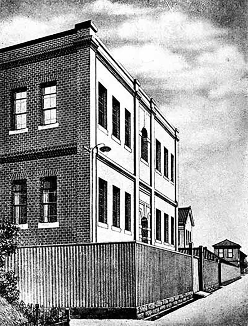
Nikon began as Nippon Kogaku K.K. (Japan Optical Industries Co., Ltd.) in July 1917 as a domestic production company of optical instruments such as rangefinders and in later years, microscopes.
Glass research facilities are built for the study of optical glass production.
Glass research facilities are built for the study of optical glass production.
Jules Bordet, the Belgian immunologist to help define the body’s immune system.
Jules Bordet, the Belgian immunologist to help define the body’s immune system.
It was discovered that the body’s immune system is capable of locating and neutralizing pathogens that have invaded the body. This system works together with immune substances called antibodies to suppress and eliminate these pathogens.
In 1919, Jules Bordet was awarded the Nobel Prize in Physiology or Medicine for his work in learning how these systems function. Microscopes were employed to observe these tiny components together with bacteria in the blood, and to help clarify how the system works. This discovery is the foundation of modern infectious disease treatment and preventative medicine.
The JOICO Microscope is the first of Nikon's microscope technology designed and manufactured in-house.
The JOICO Microscope is the first of Nikon's microscope technology designed and manufactured in-house.
The JOICO microscope is revolutionary at the time, providing a maximum magnification of 765X. The product name “JOICO” is an acronym of the Japan Optical Industry Company, the English name of the company at that time.
The physician who helped transform blood transfusions into a safe treatment and saved many lives.
The physician who helped transform blood transfusions into a safe treatment and saved many lives.
In 1900, Austrian physician Karl Landsteiner discovered that there are different types of human blood.
This identification of blood types transformed blood transfusions from being dangerous into a safe and viable treatment. This discovery saved many lives by administering blood transfusions to patients who had lost blood during surgery or in accidents. This achievement was highly praised in the medical community, and he was awarded the Nobel Prize in Physiology or Medicine in 1930.
One of the greatest medical discoveries of the 20th century, this discovery has led to saving countless lives even to present day.
One of the greatest medical discoveries of the 20th century, this discovery has led to saving countless lives even to present day.
The discovery and practical application of the antibiotic penicillin was one of the most important advances in medicine of the 20th century. Discovered by chance in 1928 from mold, penicillin was a groundbreaking drug that could be used to treat bacterial infections. At the time, many people lost their lives due to pneumonia and wound infections, and this discovery opened up the possibility of saving many lives.
For this achievement, Sir Alexander Fleming, Ernst Boris Chain, and Sir Howard Florey were awarded the Nobel Prize in Physiology or Medicine in 1945.
Today, it has become an indispensable drug for preventing and treating severe infection.
This groundbreaking microscope technology made transparent cells and microorganisms observable.
This groundbreaking microscope technology made transparent cells and microorganisms observable.
Phase contrast technology plays an important role in examining the characteristics of cancer cells, efficacy of drugs, food quality inspection and countless other microscopy applications.
This invention earned Fritz Zernike the Nobel Prize in Physics.
The model SM stereo microscope was the only product of its kind featuring a mechanism to prevent focus drift during zoom. It was also one of the first microscopes to offer epi-illumination capabilities.
The model SM stereo microscope was the only product of its kind featuring a mechanism to prevent focus drift during zoom. It was also one of the first microscopes to offer epi-illumination capabilities.
The model SM stereo microscope enables three-dimensional viewing with both eyes and featured a built-in three-step magnification optical system. Even today, stereoscopic microscopes are used not only for dissection and cultivation, but also for fine work in factories.
This small, lightweight, portable microscope, about the same size as a 35mm camera, with performance comparable to that of far larger high-end microscopes of the time.
This small, lightweight, portable microscope, about the same size as a 35mm camera, with performance comparable to that of far larger high-end microscopes of the time.
Nikon’s first inverted microscope with the objective lens located below the stage, made it suitable for higher magnification observations and greater working distances for biological and metallurgy applications.
Nikon’s first inverted microscope with the objective lens located below the stage, made it suitable for higher magnification observations and greater working distances for biological and metallurgy applications.
Albert Claude published his pioneering research on cell organelles, which was subsequently improved upon by many researchers.
Albert Claude published his pioneering research on cell organelles, which was subsequently improved upon by many researchers.
The CF technology is a revolutionary optical system that was created to provide a new expanded outlook for microscopes.
The CF technology is a revolutionary optical system that was created to provide a new expanded outlook for microscopes.
Nikon introduces the Chromatic Aberration Free (CF) system, which was developed with a completely new design concept and independently corrected chromatic aberrations of the objective lens and eyepiece. This technology was recognized as “the first innovation in 100 years.”
With the new CF technology, Nikon launches the high-grade Microphoto V series (three devices: Biophot, Metaphot, and Fluophot).
The Diaphot TMD, especially suited for the new advances in the in vitro fertilization (IVF) markets
The Diaphot TMD, especially suited for the new advances in the in vitro fertilization (IVF) markets
Microscopes developed for in vitro fertilization (IVF) are an important solution to the declining birthrate. The Diaphot TMD made a major contribution to the birth of the first baby through IVF in the United States in 1981. Since then, Nikon has been playing an important role in advancing IVF technology, helping to provide new hope to many couples.
In 2010, Robert G. Edwards, who established the IVF technique, was awarded the Nobel Prize in Physiology or Medicine. IVF is a medical technology that enables pregnancy by returning an egg fertilized outside the body with sperm to the mother’s womb, and it also contributes to the prevention of genetic diseases and the elucidation of the causes of infertility.
The nerve growth factor is a substance that develops and maintains the health of nervous tissue and the brain.
The nerve growth factor is a substance that develops and maintains the health of nervous tissue and the brain.
In the 1950s, an important substance that helps our nerve cells grow and survive is discovered. This substance is called "nerve growth factor" and function of repairing damaged nerves and extending the lifespan of nerve cells. This important discovery shed light on how the nervous system develops and is maintained, and was awarded the Nobel Prize in Physiology or Medicine in 1986. Today, it has become essential basic knowledge for research into the treatment of neurological diseases, and is used, for example, in research into treatments for Alzheimer's disease and spinal cord injuries.
Nikon’s inverted microscope is carried into orbit aboard the Space Shuttle Endeavour.
Nikon’s inverted microscope is carried into orbit aboard the Space Shuttle Endeavour.
In 1982, Nikon received an order from NASDA (National Space Development Agency of Japan, currently the Japan Aerospace Exploration Agency (JAXA)) for an inverted biological microscope to be used aboard the space shuttle. It was delivered in late 1985 and was carried into orbit on the Space Shuttle Endeavour in 1992. It was primarily used for material experiments in outer space.
Nikon’s first confocal microscope, the RCM8000 is introduced.
Nikon’s first confocal microscope, the RCM8000 is introduced.
Nikon launches its first real-time laser confocal microscope, the RCM8000. This microscope was capable of capturing images of specific areas of thick specimens with extremely high clarity which was difficult to do with regular optical microscopes.
Edward B. Lewis et al. identifies specific genes that help shape living organisms.
Edward B. Lewis et al. identifies specific genes that help shape living organisms.
This team were responsible identifying genes that help guide the proper body formation of an organism.
Working with fruit flies, the team discovered a specific gene that controls the formation of body parts. This gene plays a key role in directing where and how each part of the body is formed, from head to tail.
Even more surprising, it was revealed that this genetic mechanism works in almost the same in flies as it does in vertebrates, such as mice and humans. This discovery has greatly advanced our understanding of human fetal development and congenital diseases.
Edward B. Lewis, Christiane Nüsslein-Volhard, and Eric F. Wieschaus were awarded the Nobel Prize in Physiology or Medicine for their discovery.
Nikon develops an innovative microscope optical system which greatly extends the degree of freedom for optical design from 45mm to 60mm providing a greater enhanced field of view. Nikon introduces the E800, the first microscope system to employ this new technology.
Nikon develops an innovative microscope optical system which greatly extends the degree of freedom for optical design from 45mm to 60mm providing a greater enhanced field of view. Nikon introduces the E800, the first microscope system to employ this new technology.
The distance from the mounting surface of the objective lens to the observation target (same focal length) was extended to 60mm from the existing 45mm. This greatly increased the degree of freedom for optical design and achieved the world's first 0.5x objective lens, realizing observation of an ultra-wide field of view with a diameter of 50mm. The ECLIPSE E800 was an innovative microscope that employed this system.
This discovery played a key role in solving the mystery of how proteins reach the correct location within cells.
This discovery played a key role in solving the mystery of how proteins reach the correct location within cells.
Our cells contain many different types of proteins. These proteins maintain our body's normal functions by working in specific locations within the cells.
It was learned that proteins carry a small marker called a “signal peptide,” which guides them to their proper destinations within the cell.
Understanding this mechanism has helped scientists learn more about various diseases that result from disruptions in protein transport.
This breakthrough earned Dr. Günter Blobel the Nobel Prize in Physiology or Medicine.
The flagship inverted microscope system “ECLIPSE TE2000” is announced, combining the new CFI optical technology with expanded capabilities for new digital imaging developments.
The flagship inverted microscope system “ECLIPSE TE2000” is announced, combining the new CFI optical technology with expanded capabilities for new digital imaging developments.
The ECLIPSE TE2000 Inverted Microscope was a completely new generation of microscope designed to be compatible with the then new digital imaging technologies. This new system offered excellent flexibility with multiple ports to allow increased capabilities within the system. This microscope quickly became Nikon’s flagship microscope.
The COOLSCOPE is introduced as Nikon’s first of its kind pioneer in the digitalization and automation of microscopes.
The COOLSCOPE is introduced as Nikon’s first of its kind pioneer in the digitalization and automation of microscopes.
The COOLSCOPE is introduced as Nikon’s first of its kind pioneer in the digitalization and automation of microscopes.
The discovery of this mechanism uncovered the mystery of our sense of smell.
The discovery of this mechanism uncovered the mystery of our sense of smell.
This team was credited for uncovering how the brain identifies the many different smells we perceive.
At the back of the interior of the human nose, there are approximately 1,000 types of specialized receptors, each detecting specific odor molecules. These receptors work in various combinations, allowing us to distinguish around one trillion different scents.
This discovery marked a major breakthrough in understanding how the brain processes external information and how our sense of smell works. In particular, the revelation of how neurons selectively recognize specific types of information has significantly influenced research in other sensory systems as well.
Richard Axel and Linda B. Buck were awarded the Nobel Prize in Physiology or Medicine.
The BioStation CT cell culture observation system is introduced which combined robotics designed by Nikon for other applications with biological microscope technology to provide a system that could automatically conduct experiments on multiple samples simultaneously.
The BioStation CT cell culture observation system is introduced which combined robotics designed by Nikon for other applications with biological microscope technology to provide a system that could automatically conduct experiments on multiple samples simultaneously.
A groundbreaking device that combines a microscope with a device for maintaining constant temperature and humidity for cell culture, allowing cells to be observed with reduced stress. It has been introduced in cutting-edge laboratories around the world, including the laboratory of Professor Shinya Yamanaka of Kyoto University, who was the first in the world to successfully create iPS cells. With its automated observation function, it can be said to be a pioneering product in laboratory automation.
Launch of the ECLIPSE Ti-E inverted microscope equipped with PFS
Launch of the ECLIPSE Ti-E inverted microscope equipped with PFS
Equipped with PFS (Perfect Focus System) and a high-speed motorization capability, this system meets the needs of long-term live cell imaging in biological, medical, and pharmaceutical science research.
PFS uses unique optical technology to automatically track any Z-axis plane while detecting the interface with the slide glass. The built-in linear encoder and high-speed active feedback quickly correct any focus deviation with high precision. Even during long-term acquisition of complex continuous images, it always provides focused and reliable images. This technology was recognized as one of the TOP INNOVATIONS of 2008 in The Scientist magazine.
The discovery and development of green fluorescent protein (GFP) revolutionized fluorescence imaging in microscopy.
The discovery and development of green fluorescent protein (GFP) revolutionized fluorescence imaging in microscopy.
Discovered in the jellyfish Aequorea victoria, ""green fluorescent protein (GFP)"" is a special protein that glows bright green when exposed to ultraviolet light. It was learned that this property can be used to track the movement of specific proteins inside living cells. This discovery made it possible to directly observe life phenomena that were previously invisible, such as the proliferation process of cancer cells and the functioning of neurons in the brain.
The importance of GFP has been widely recognized in the scientific community as a major advancement in imaging technology, and in 2008 its discoverers were awarded the Nobel Prize in Chemistry.
Nano Crystal Coat technology is applied to Nikon optics for non-reflective capabilities that enhanced image clarity significantly.
Nano Crystal Coat technology is applied to Nikon optics for non-reflective capabilities that enhanced image clarity significantly.
The lens anti-reflection coating called "Nano Crystal Coat" was originally developed for semiconductor manufacturing equipment, which required extraordinary precision and performance. Nikon quickly recognized that this technology would also have benefits to scientists imaging through microscope objective lenses. This advancement made it possible to develop objective lenses with excellent light transmittance and reduced light reflection across a wide range from ultraviolet to near infrared.
N-SIM and N-STORM super-resolution microscopes are introduced Microscopes for the first time ever are able to make observations beyond the resolving capabilities of optical lenses.
N-SIM and N-STORM super-resolution microscopes are introduced Microscopes for the first time ever are able to make observations beyond the resolving capabilities of optical lenses.
Super-resolution microscopy is an innovative technology that has made it possible to observe extremely small structures inside cells that could not be seen with conventional optical microscopes. While conventional optical microscopes could not observe objects smaller than 200 nanometers, this technology enables observation down to 20 nanometers (about 1/5,000th the thickness of a human hair).
Nikon’s super-resolution microscope system enables observation of the fine structure of living cells and molecular level observation with a resolution far exceeding the limits of conventional optical microscopes. Furthermore, it enables high-speed acquisition and observation at the single-molecule level.
This technology allows real-time observation of phenomena occurring within living cells, and has made significant contributions to elucidating the movement of proteins and the process of cell division in particular.
The development of this groundbreaking super-resolved fluorescence microscopy led to Eric Betzig, Stefan Hell, and William Moerner being awarded the Nobel Prize in Chemistry in 2014. Currently, super-resolution microscopes are used in research institutions around the world, helping to develop new treatments and elucidate biological phenomena.
This discovery greatly expanded the possibilities for regenerative medicine.
This discovery greatly expanded the possibilities for regenerative medicine.
Once our cells have a specific role (skin, muscle, nerves, etc.), they usually do not transform into another type of cell.
However, in 2006, a groundbreaking discovery was made that overturned this conventional wisdom. It was proven that by introducing specific genes into mature cells, they can be restored to a young state and changed into any type of cell.
These “rejuvenated cells” are called “iPS cells.”
This discovery has opened the possibility of creating necessary tissues from a patient's own cells, which is being applied to the treatment of intractable eye diseases and retinal regenerative medicine, for example.
In 2012, Sir John B. Gurdon and Shinya Yamanaka were awarded the Nobel Prize in Physiology or Medicine for their discovery.
Uncovering the intricate transport mechanisms of the microscopic world.
Uncovering the intricate transport mechanisms of the microscopic world.
There is an elaborate mechanism for transporting substances within cells. This mechanism is called “vesicular transport,” and it packs proteins and other substances into small sacs (organelles) and delivers them to the necessary locations.
Three scientists who have made a major contribution to elucidating this mechanism are James E. Rothman, Randy W. Schekman, and Thomas C. Südhof. They discovered the genes necessary for intracellular transport, and elucidated the mechanisms of the necessary proteins and the mechanism by which nerve cells release neurotransmitters at precise times. In recognition of this research, they were awarded the Nobel Prize in Physiology or Medicine in 2013.
The discovery of “place” cells that work like a GPS within our brains.
The discovery of “place” cells that work like a GPS within our brains.
How are we able to remember which way to go? The answer to this question was clarified by the discovery of the brain's “location-positioning system.”
In 1971, John O'Keefe discovered ""place-specific cells"" in the hippocampus of the brain. These cells react when we become aware of our current location. Then, in 2005, May-Britt Moser and Edvard Moser discovered ""grid cells,"" which record even more detailed location information. They discovered that these two types of cells work together like a GPS.
This discovery was awarded the Nobel Prize in Physiology or Medicine in 2014, and is now being applied to research into dementia and Alzheimer's disease.
Autophagy is an intracellular recycling system that breaks down and reuses substances within the cell.
Autophagy is an intracellular recycling system that breaks down and reuses substances within the cell.
Our cells have a mechanism called “autophagy” that breaks down and reuses unnecessary substances. This was discovered in the 1960s, but its details remained a mystery for many years.
Yoshinori Okuma learned the mechanism of autophagy at the genetic level through experiments using yeast.
Since then, research has progressed and it has become clear that abnormalities in autophagy are related to various diseases such as Parkinson's disease, which expresses itself as abnormal protein accumulation. It is thought that impaired autophagy function might be one of the causes for this disease. In addition, development of new therapeutic drugs that target autophagy is progressing, and it is expected that they will be applied to the treatment of cancer and neurological diseases.
For his work, Ohsumi Yoshinori was awarded the Nobel Prize in Physiology or Medicine in 2016.
Research into “releasing the brakes” of immune cells.
Research into “releasing the brakes” of immune cells.
In 1992, Honjo Tasuku discovered a molecule which is involved in our body's immune system. Normally, immune cells attack cancer cells to protect us, but this molecule known as PD-1 inhibits this function. Honjo discovered that by “releasing the brakes”, immune cells can once again fight cancer cells.
The therapeutic drug developed based on this discovery was the first of its kind in the world to be approved, in 2014. It is currently used to treat a variety of cancers, including malignant melanoma and lung cancer.
In 2018, Honjo Tasuku and James P. Allison were awarded the Nobel Prize in Physiology or Medicine for their “groundbreaking discoveries in cancer treatment.”
Supporting users in a cutting-edge biotechnology facility.
Supporting users in a cutting-edge biotechnology facility.
To provide wide range of support for individual users in areas such as basic drug discovery research, candidate drug screening, and optimization of cell culture conditions, Nikon opened the Nikon BioImaging Lab in 2019 in Boston, which is a biocluster bringing together pharmaceutical companies and bio ventures. In 2021, Nikon also opened a Nikon BioImaging Lab at Shonan iPark in Japan, followed by another at the Leiden Bio Science Park (LBSP) in the Netherlands. By utilizing Nikon’s live-cell imaging and image analysis technologies, these labs contribute to the efficient development of pharmaceuticals.
Technological advances incorporated to yield wide-range, high-resolution, ultra-high-speed imaging is realized.
Technological advances incorporated to yield wide-range, high-resolution, ultra-high-speed imaging is realized.
The AX and AX R confocal microscopes were developed to meet the requirements for observing the minute structures of large specimens while they are still alive, and to study and analyze their instantaneous reactions and changes. Cross-sectional images can be obtained with high contrast, and specimens can be observed in three dimensions by combining multiple images. The high-performance detectors and scanners enable wide-range, high-resolution, ultra-fast imaging. They contribute to researchers understanding life phenomena more efficiently and in greater detail.
Supporting pathologists with unique design and functionality.
Supporting pathologists with unique design and functionality.
Despite being a microscope, the ECLIPSE Ui features a unique design that does not require an eyepiece. In addition to improving the posture of pathologists during observation, it also enables the sharing of observed images on a computer display. This leads to reducing the issues and fulfilling the needs of pathology sites, such as the shortage of pathologists and the accumulation of fatigue on users due to long hours of observation, and contributes to improving the workflow of pathology diagnoses.
AI automation incorporated into automated microscopy system to streamline studies in biomedical research and drug discovery.
AI automation incorporated into automated microscopy system to streamline studies in biomedical research and drug discovery.
Using AI, the process from image acquisition to analysis has been largely automated. This makes microscope operation more efficient, allowing users to focus on more creative activities such as analysis and observation. The system also maintains hardware expandability, contributing to the efficiency of research and development in drug discovery.
The ECLIPSE Ti2-I motorized inverted microscope for is introduced as an automated configuration to improve the efficiency of infertility treatment.
The ECLIPSE Ti2-I motorized inverted microscope for is introduced as an automated configuration to improve the efficiency of infertility treatment.
As the number of infertility treatments increases, so does the demand for higher accuracy and efficiency of treatments. In response, Nikon developed a product specialized for ICSI using a microscope. By automating the settings, the number of steps required to operate the microscope has been significantly reduced, contributing to improvements in the efficiency of ICSI.
Note:
- The products introduced here are based on their Japanese release dates, while these products are sold globally. Please note that product availability, release timing, and primary intended use may vary depending on your region/country.
- The scientific discoveries presented here are based on the year the Nobel Prize was awarded, not the actual year of the discovery. Please note that this may differ from the actual year the discovery was made.


