Nikon Europe B.V. | Europe & Africa
- pt Change Region
- Global Site
The services offered by the Nikon BioImaging Lab -Shonan- is unlike a traditional core facility, our services don’t stop at assisted equipment use, but include up to full service imaging, and analysis.
Not sure if your needs fit strictly within one of the below service options? There’s no need to worry, we are flexible in designing a relationship to provide the services you need, and not the ones you don’t. These are simply guidelines to help explain the capabilities of the Nikon BioImaging Lab, and avenues for engaging with us.
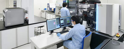
The information on these pages applies to the Nikon BioImaging Lab in Shonan, Japan. There are also laboratories in Leiden, The Netherlands and Cambridge, Massachusetts, USA. For more information, please contact the facility nearest to your location.
Our pilot program enables you to evaluate whether the Nikon BioImaging Lab’s contract imaging services are a suitable for your research. Collaborate with Nikon's expert staff to explore your research requirements and help determine how your project will function in practice. Please contact us for more information on this trial service.
In the drug discovery field, cells and biological tissues are used for evaluating the efficacy and safety of drug candidates under conditions aimed at recapitulating the physiological environment. Before administering a new investigational drug to humans to confirm its efficacy and toxicity, an enormous amount of time and cost is required in order to reproducibly carry out such tests.
In contrast, an organ chip can mimic an organ in-vivo system with physiological functions by culturing cells derived from the organ in a micro-fluidic device on the chip. While this is a revolutionary technology that can be utilized to evaluate the efficacy and toxicity of candidate compounds with high accuracy and efficiency, advanced techniques are required to acquire images of organ chips with complex structures, select analysis conditions, and acquire quantitative data. Nikon has established the optimum conditions for imaging and analysis for each organ-on-a-chip and the specific aim of each experiment, in collaboration with major organ-on-a-chip manufacturers.
Nikon provides “Organ-on-a-Chip Imaging and Analysis contract services” that capture and analyze organ chip images in the most suitable manner for the purpose of research and development relating to drug discovery, based on commissions from each customer, at Nikon BioImaging Labs.
*1 This service targets the OoC (Organ-on-a-Chip) samples of seeded, stained, and fixed cells.
| Available Samples at NBIL |
|---|
| Emulate Chips |
| Emulate PODs |
| Mimetas Organoplates |
| AIM Biotech Chips |
| Nortis Chips |
| Tissuse Chips |
Please contact us for service costs and details.
Drop off your samples and leave the imaging to Nikon. We’ll send back the imaging data in your preferred format. This option is best for customers who want to do their own image analysis and quantification, but don’t have sufficient time, personnel, and/or equipment availability for desired type of imaging. You choose the level of culturing service that makes sense for the chosen service(s).
We perform all qualitative and quantitative analyses on images and report complete results. This service applies to both images acquired at the Nikon BioImaging Lab and elsewhere using other microscope systems, including those produced by other manufacturers. This service may even be provided remotely, expanding its availability beyond the Shonan and Tokyo area. We can even work with our Development section to craft custom analysis solutions for demanding applications, including solutions based on artificial intelligence/machine learning.
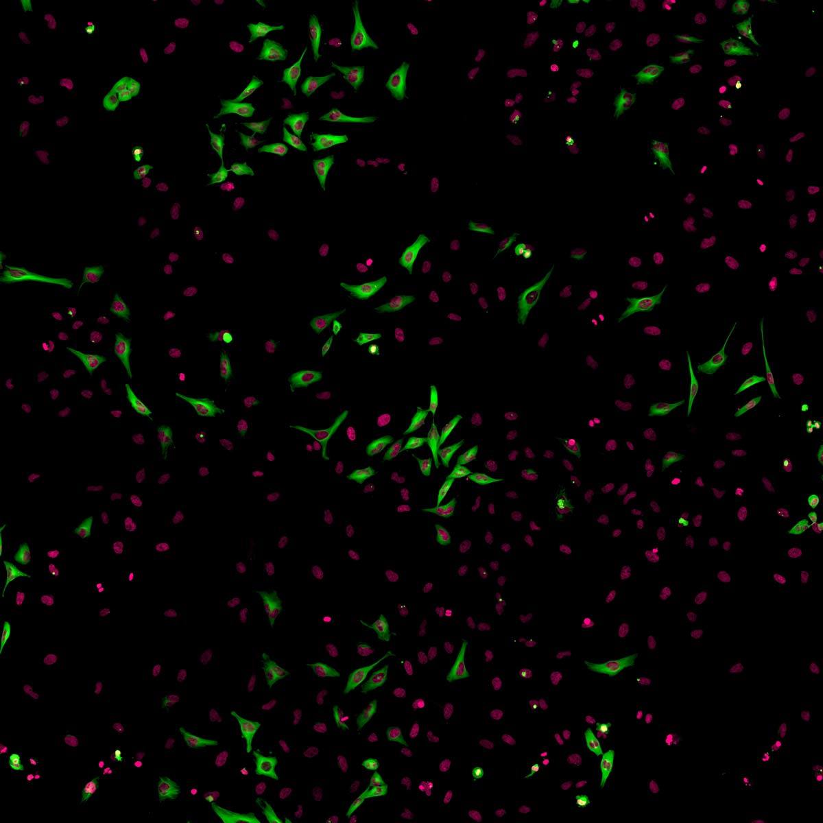
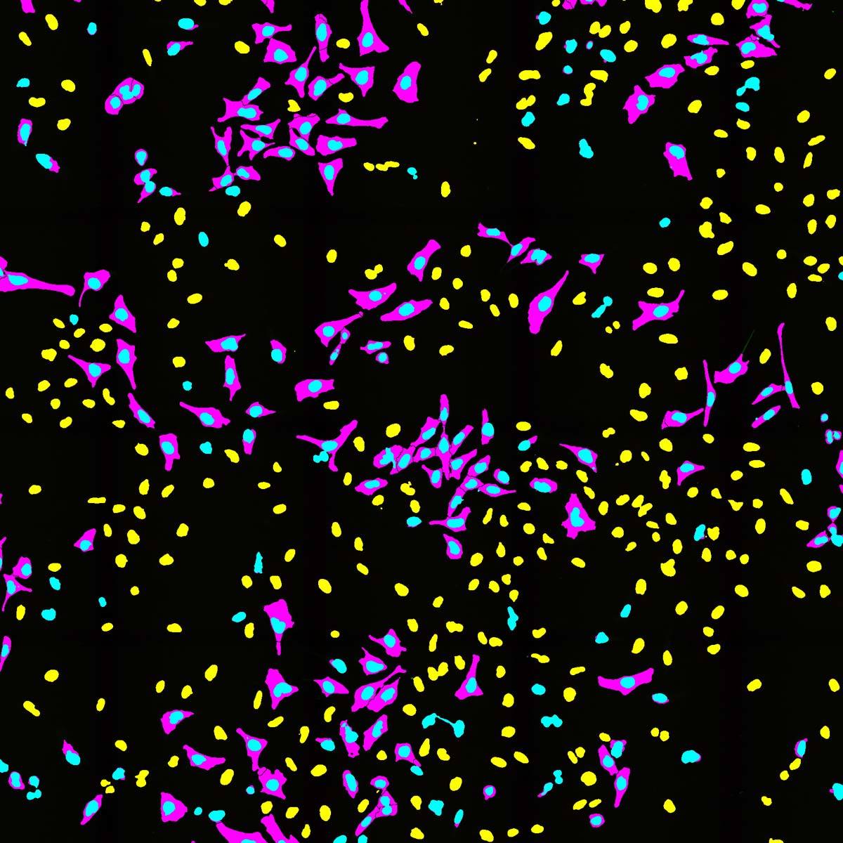
A ferramenta de classificação de células segmenta e classifica as amostras em categorias para contagem e medição
Experienced and semi-experienced users can bring samples and have us walk them through the acquisition and analysis process. We can even provide microscopy education, training, and consulting services to help maximize the effectiveness of your group with image-based research more generally. Once users have had sufficient time to familiarize themselves with the equipment, we can transition to an independent equipment use model as detailed in the next section.
Experienced microscopy users can rent time on our instrumentation at a simple hourly rate, similar to a traditional imaging core facility. Equipment use includes both imaging with our various microscope systems, as well as the use of our NIS-Elements software for image analysis and reporting applications. It is also possible to begin with an assisted equipment use arrangement and to transition to independent use once you are comfortable doing so.


