Nikon Instruments Inc. | Americas
- fr Change Region
- Global Site
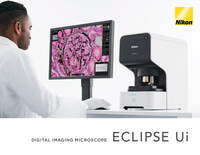
Nikon’s new ECLIPSE Ui Digital Upright Microscope provides accurate microscopy-based observation imaging. View and share high quality images in real time with easy-to-use software for a more streamlined workflow.
This product is currently available in the United States only. Please contact your local dealer for more information on this product in your country.
The ECLIPSE Ui’s image quality is backed by Nikon's renowned imaging technology — clear color reproducibility without negative influence from ambient light. Furthermore, the elimination of conventional eyepieces greatly reduces operator eye fatigue allowing the user to work effortlessly for longer periods of time.
The internal PC provides all the necessary functions and applications.
It is no longer necessary to sit for hours looking through microscope eyepieces. The images are shared onscreen, suitable for two or more people to discuss the samples.
Nikon CFI Plan Fluor Series Objective Lenses
The ECLIPSE Ui is equipped with Nikon’s exceptional CFI Plan Fluor Series objectives. These lenses are acclaimed for their high standard: they have superior optical transmission and high numerical aperture, yielding better resolved images.
PAM staining of membranous nephropathy (40X)
The Nikon ECLIPSE UI’s unique design produces high-quality images that are easy to observe while afterimages are minimized, yielding better color reproducibility and allowing smooth and fast scrolling with adjustable brightness and color tone.
Samples can be completely observed. And the positions, cell distributions and other factors of samples on slide can also be quickly checked*1.
In addition, the digital zoom function allows you to zoom in on the macro images for reliable observation.
*1 Macro images are intended to put samples in place. For diagnostic purposes, be sure to use the Live Image (micro image) with a 4x or higher objective lens.
*2 For details on image capture in the high-quality mode, refer to the Specifications.
The ECLIPSE Ui is operational in 2.5 seconds after loading a sample providing the user an immediate viewable image. Digital sample images can be observed live. Furthermore, magnification changes and X-Y movements can be quickly adjusted. It is also equipped with a macro-imaging function and other sample-oriented applications.
A prepared slide can be placed on the unit with only one hand for quick and efficient slide exchange.
Samples are loaded at the touch of a button and appear on the display monitor in 2.5 seconds.
A sample is captured as a macro-image, thereby allowing the user to quickly identify the region of interest to be observed. That site can also be selected and preset so that it can be recalled with just one click. The macro-image displayed may also be saved as required.
A Live Image can be turned 90-degrees for reliable observation.
Thick samples can be easily observed using the scroll wheel or focus button
The system is designed to meet various use cases, such as thick sample observation and imaging.
The Z-focus can be quickly repositioned for thick or undulating samples by using the mouse scroll wheel or onscreen focus button.
The stage can be successively shifted at a constant rate to enable convenient, multi-field observation. There are six speed settings for a wide range of scrolling applications.
The ECLIPSE Ui system is equipped with a GUI for easy identification and for efficient observation tasks.
The functions needed to observe sample images are arranged in a user-friendly way to allow one to simultaneously view micro- and macro-images in real time.
Digital zoom-in/out available.
Equipped with the auto-focus function or focus adjustment is also possible with the onscreen buttons or the mouse wheel.
Exposure and aperture control are easily adjusted by dragging the slide bars onscreen.
Shades and contrasts can be quickly and easily adjusted to the user’s preferences.
Areas of interest can be annotated and point-to-point measurements can be made within the displayed image.
The functions unique to the ECLIPSE Ui, such as tracing of already observed field of views and automatic alignment serve to observe samples more objectively and consistently.
Two samples of a tissue, differently stained, can be visualized side by side at a time. Comparative observations are now made easy and help pathologists double-check effectively. The two samples are automatically aligned using this feature. You can also readily navigate between the various target points with a single click.
H&E-stained sample and low-contrasted stained sample are automatically aligned, and their registered observation points are also automatically photographed in sequence.
Up to 10 sample images can be displayed all at once in the multi-split-screen for each registered observation point. The same observation points of multiple stained samples can be reviewed in grid layouts, enabling efficient comparative observation*4.
*4 Simultaneous comparative observations with multiple stained samples, such as ER-stained, PgR-stained and Ki67-immunostained.
Trace Display shows which areas in the sample were previously viewed – indicated by a slight shading in the macro image. This helps more efficient observation and contributes to preventing oversight of critical areas.
The ECLIPSE Ui system is equipped with three modes: routine specimen observation, research*3/education*3, and data sharing. Users can easily select their preferred imaging quality and speed.
In this mode, cytoscreening is supported. Sample images displayed live on the monitor screen can be used for routine pathological image observation and successive observation. Captured images can be transferred to other pathological systems for later viewing.
Data (sample images, observation spots, etc.) to share or discuss are saved in an external storage*5. This data can be utilized for relevant studies and education.
The system can be controlled remotely (users on contract) allowing the user to observe real time imagery from anywhere*6.
*3: Not for use in diagnostic procedures.
*5: Separately sold.
*6: Separate contract must be concluded for using the Remote mode. For communication environment, contact us.
The small footprint of the body measures 422 mm in height, 233 mm in width and 427 mm in depth. The microscope can be readily and easily set up and does not require a dark room as ambient light does not affect the image.
Bar code and 2D code (QR code) are easily read, displayed and saved, reducing the chance of samples becoming mixed-up.
3712 x 3712, above 1 fps
There are two types of image output: LIVE for immediate observation and evaluation, and High-Quality*7 for saving and storage.
LIVE (Observation mode)
1080 x 1080, above 30 fps
High-Quality*7 (Capture mode)
3712 x 3712, above 1 fps
*7 Saved images cannot be used for diagnostic purposes.
Users (on contract) in remote locations may access and operate the system to allow for shared medical treatment discussion, collaboration and immediate and accurate observation.
*8 For this Remote mode functionality, a separate contract must be used. Contact Nikon for communication environment information.
Sample is loaded by the Remote user.
The same image is reviewed both by the user on contract and Remote user.
The system is operated both by the user on contract and Remote user (for focusing, positioning, magnification adjustment, etc.).
Sample is removed by the Remote user.
Frozen sections can be quickly observed from any remote location with the ECLIPSE Ui.