Nikon Instruments Inc. | Americas
- fr Change Region
- Global Site
Director of the Department of Reproductive Medicine, Center for Regenerative Medicine, National Center for Child Health and Development
The National Center for Child Health and Development, where I work, is a medical facility that conducts medical care related to growth diseases*, promotes greater understanding of such diseases and undertakes research into treatments for them. In this field where clinical practice and research are so closely linked, I think this facility is the most advanced in Japan. I am conducting research on human pluripotent stem cells employing regenerative medicine and related technology in order to contribute to the establishment of treatment and diagnostic methods for children with intractable diseases. I have worked as an obstetrician in a clinical setting myself, so I always keep it in mind to utilize the results of research to improve the quality of life of patients. Developing a “mini-gut” is a part of this.'
*Growth diseases: Diseases that occur during the human life cycle from fertilization/pregnancy to fetal, neonatal, infancy, school-age, adolescence, and adulthood.
Simply put, a mini gut is a “miniature gut” made from human pluripotent stem cells. It is a three-dimensional structure, with a size of about 1 cm to 2 cm. Unlike conventional intestinal organoids*, a mini gut consists of not only intestinal mucosal epithelial tissue, but also the muscles and nerves of the submucosal tissue. It also functions as a small intestine, including absorption and metabolism of nutrients, peristalsis, response to drugs and immune function. Furthermore, the mucosal epithelium faces outwards as if the actual small intestine had been turned inside out. For this reason, it is very easy to observe reactions and effects due to the administration of drugs.
*Organoids: Tissues that mimic bodily organs. Using stem cells and iPS cells, they are formed in vitro, including the three-dimensional structure.
Mini gut, about 1 cm in size
In addition to digestion and absorption, the small intestine also helps protect the human body from bacteria and viruses and promotes the secretion of hormones such as insulin. While it is an important organ that greatly contributes to people’s quality of life, it possesses a complex structure and diverse functions, so it is susceptible to many diseases, for many of which no treatment or even diagnostic methods have been discovered.
Many diseases that occur during childhood are related to the process by which organs are formed. So this made it difficult to advance our understanding unless we could comprehend the entire organ. The mini-gut is able to live in a culture medium for more than one year after production. This makes it possible to conduct longer-term observation, research, and evaluation, and to explore the mechanisms of various diseases as well as effective treatments and diagnostic methods. With the advancement of technology, new intestinal diseases have been discovered, while patients to be treated are increasingly of a younger age. So, in order to effectively understand diseases and connect them to diagnosis and treatment, human intestinal models will be increasingly required in the future. I strongly hope that the mini-gut can be utilized for such research.
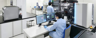
A miniature organ such as the mini-gut is composed with three-dimensional tissues of multiple cells, and it exhibits the same functions as real organs in the body. While the more sophisticated the functions of a miniature organ are, the more important it becomes to evaluate them accurately. For the mini-gut, it is essential to acquire images stereoscopically and at high resolution. Therefore, we asked the Nikon BioImaging Lab to perform the imaging that we usually handle in our laboratory. As a result, high-resolution images that we had never seen before were obtained. I was surprised at how high the resolution was and felt strongly that I could take another step forward in my research.
Confocal 3D imaging of a mini-gut
Cross-sectional images of a mini-gut
In the case of the mini-gut, assessment of movement was also a major challenge. For example, it is possible to convert the movement of a single myocardial cell into an electrical signal and evaluate it. However, in a mini-gut, the entire tissue moves in an undulating motion. These peristalsis movements couldn’t be evaluated before. We had filmed a video ourselves and asked various establishments to evaluate the movement recorded in it, but quantitative evaluation was not possible. When we finally entrusted the work to the Nikon BioImaging Lab, the peristaltic movements were successfully quantified by using analysis software* developed by Nikon.
*Imaging software NIS-Elements. For more details, click here.
Peristaltic movements of a mini-gut
In order to accurately evaluate a mini-gut, it is crucial to acquire three-dimensional rather than two-dimensional images. It is necessary to capture the localization of proteins that cause diseases with high resolution, and also essential to capture the complex movements of living tissues over time. Only when all of these conditions are satisfied and the software for analyzing such images is available, does accurate evaluation of the mini-gut become possible. Moreover, as its usage increases, the application fields will expand. Mini-gut research and microscopic imaging techniques are inseparable.
I would like to observe certain sizes of mini-gut over periods of time and discover their various functions. Especially, if we could capture the progression of a reproduced disease at a speed close to real time, we might be able to find ways of improving the areas where the movement function is declining. For example, it could be utilized in drug discovery screening for treating diseases. For that reason, I would be even happier if there was a function that could automatically calculate the quantitative evaluation results of the captured video. Though I think we might possibly be asking too much in many ways.
Plus, it would be great if we could acquire high-resolution images with even more depth than those currently achieved, allowing us to analyze them with even higher accuracy. For drug discovery applications it is extremely important to have a method for evaluating whether a drug is effective or not. To that end, with the major premise of taking clear, beautiful images, I believe that it will be very important to capture the target more three-dimensionally in real time, and to analyze and evaluate the target measurement data as quickly and accurately as possible.
After I published my paper on the mini-gut, I was surprised to learn that people from a diverse range of industries and fields were showing great interest in my research. They came from pharmaceutical companies, as well as from general consumer goods business fields such as foodstuffs, supplements, and sanitary products. This wide variety gave me a sense of the great potential for future development. However, my original mission is to utilize the mini-gut to contribute to the diagnosis and treatment of intestinal disease in children. Even if just one treatment is effective, I want to contribute to it as rapidly as I can. Whether it’s through drug discovery, the establishment of diagnostic methods, or prevention, in whatever form it takes, I’d like to contribute as soon as possible.
Talking about the future, just one possible example, if we can develop a drug that causes the intestines to lengthen, it could help treat children who are suffering from congenital short intestines.
Improvement for them could be achieved using only a single medicine, without them undergoing what is major surgery for a small body. In order to realize such a future, I think it is vitally important to collaborate not only with researchers but also with clinicians who face patients every day, pharmaceutical companies, and optical manufacturers that develop microscopes. I sincerely believe that the opportunities for Nikon to help us in our task will only continue to increase.


