![](https://downloads.microscope.healthcare.nikon.com/production/imager/mastheads/2210/LV100ND-POL-DS-Masthead_f076438097b2d2d85c6c3ef04742411e.jpg)
Los microscopios de luz polarizada pueden medir las propiedades del amianto, como los índices de refracción, la birrefringencia, el retraso de fases, los ángulos de extinción, el pleocroísmo y los signos de elongación, lo que ayuda a identificar el amianto. LV100ND POL / DS es un microscopio de luz polarizada de alto rendimiento con accesorios que permiten la observación de manchas de dispersión de hasta 400 aumentos.
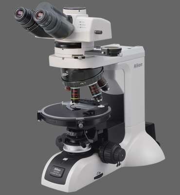
- El LV100ND POL / DS emplea óptica con altos factores de extinción, facilitando la detección y observación de propiedades ópticas de alta precisión.
- El LV100ND POL / DS permite el análisis de birrefringencia, ángulos de extinción, signos de elongación, índices de refracción, magnitudes de birrefringencia y pleocroísmo del asbesto.
- Cambiando los objetivos y los módulos PH permiten la observación de la dispersión coloreada del asbesto correspondientes a los índices de refracción del líquido de inmersión. El LV100ND POL / DS permite el análisis cuantitativo y la observación de manchas de dispersión del asbesto.
- Una platina circular graduada completamente giratoria ofrece rotación de la muestra sin problemas y el análisis preciso durante la observación de polarización.
![](https://downloads.microscope.healthcare.nikon.com/production/imager/productphotos/2165/lv100nd-pol-ds_3cac538b21a254209ea06cf584646f29.jpg 1x, https://downloads.microscope.healthcare.nikon.com/production/imager/productphotos/2165/lv100nd-pol-ds_7fc2443c96341e0fab73dbc596e4b275.jpg 2x)
Características principales
Highlights
- The LV100ND POL/DS employs optics with high extinction factors, facilitating detection and observation of high-precision optical properties.
- The LV100ND POL/DS enables analysis of birefringence, extinction angles, signs of elongation, refractive indices, birefringence magnitudes and pleochroism of asbestos.
- Changing the objectives and PH modules allows observation of asbestos dispersion colors corresponding to the refractive indices of the immersion liquid. The LV100ND POL/DS enables both quantitative analysis and dispersion staining observation of asbestos.
- A fully rotatable circular graduated stage offers trouble-free sample rotation and accurate analysis during polarization observation.
![](https://downloads.microscope.healthcare.nikon.com/production/imager/keyfeatures/LV100ND-POL-DS/2234/LV100ND-POL-Birefringence1_eab673dc52b0446b2b598a80abc7b050.jpg 1x, https://downloads.microscope.healthcare.nikon.com/production/imager/keyfeatures/LV100ND-POL-DS/2234/LV100ND-POL-Birefringence1_9b9989dbe1d056a5c35107959161a1c5.jpg 2x)
La cruz Nicols, posición diagonal
![](https://downloads.microscope.healthcare.nikon.com/production/imager/keyfeatures/LV100ND-POL-DS/2235/LV100ND-POL-Birefringence2_eab673dc52b0446b2b598a80abc7b050.jpg 1x, https://downloads.microscope.healthcare.nikon.com/production/imager/keyfeatures/LV100ND-POL-DS/2235/LV100ND-POL-Birefringence2_9b9989dbe1d056a5c35107959161a1c5.jpg 2x)
La cruz Nicols, posición de extinción
![](https://downloads.microscope.healthcare.nikon.com/production/imager/keyfeatures/LV100ND-POL-DS/2238/LV100ND-POL-Extinction-angle1_eab673dc52b0446b2b598a80abc7b050.jpg 1x, https://downloads.microscope.healthcare.nikon.com/production/imager/keyfeatures/LV100ND-POL-DS/2238/LV100ND-POL-Extinction-angle1_9b9989dbe1d056a5c35107959161a1c5.jpg 2x)
El Nicols paralelo, cara de escisión alineada con la cruz
![](https://downloads.microscope.healthcare.nikon.com/production/imager/keyfeatures/LV100ND-POL-DS/2237/LV100ND-POL-Extinction-angle2_eab673dc52b0446b2b598a80abc7b050.jpg 1x, https://downloads.microscope.healthcare.nikon.com/production/imager/keyfeatures/LV100ND-POL-DS/2237/LV100ND-POL-Extinction-angle2_9b9989dbe1d056a5c35107959161a1c5.jpg 2x)
La cruz Nicols, ángulo de extinción medido
![](https://downloads.microscope.healthcare.nikon.com/production/imager/keyfeatures/LV100ND-POL-DS/2241/LV100ND-POL-DS-elongation1_eab673dc52b0446b2b598a80abc7b050.jpg 1x, https://downloads.microscope.healthcare.nikon.com/production/imager/keyfeatures/LV100ND-POL-DS/2241/LV100ND-POL-DS-elongation1_9b9989dbe1d056a5c35107959161a1c5.jpg 2x)
Amosite positivo: Posición diagonal sustractiva
![](https://downloads.microscope.healthcare.nikon.com/production/imager/keyfeatures/LV100ND-POL-DS/2240/LV100ND-POL-DS-elongation2_eab673dc52b0446b2b598a80abc7b050.jpg 1x, https://downloads.microscope.healthcare.nikon.com/production/imager/keyfeatures/LV100ND-POL-DS/2240/LV100ND-POL-DS-elongation2_9b9989dbe1d056a5c35107959161a1c5.jpg 2x)
Crocidolita Negativo: Posición diagonal aditiva
![](https://downloads.microscope.healthcare.nikon.com/production/imager/keyfeatures/LV100ND-POL-DS/2247/LV100ND-POL-DS-refractive-index1_eab673dc52b0446b2b598a80abc7b050.jpg 1x, https://downloads.microscope.healthcare.nikon.com/production/imager/keyfeatures/LV100ND-POL-DS/2247/LV100ND-POL-DS-refractive-index1_9b9989dbe1d056a5c35107959161a1c5.jpg 2x)
La parte superior de un espécimen está enfocada
![](https://downloads.microscope.healthcare.nikon.com/production/imager/keyfeatures/LV100ND-POL-DS/2246/LV100ND-POL-DS-refractive-index2_eab673dc52b0446b2b598a80abc7b050.jpg 1x, https://downloads.microscope.healthcare.nikon.com/production/imager/keyfeatures/LV100ND-POL-DS/2246/LV100ND-POL-DS-refractive-index2_9b9989dbe1d056a5c35107959161a1c5.jpg 2x)
La parte inferior de un espécimen está enfocada
![](https://downloads.microscope.healthcare.nikon.com/production/imager/keyfeatures/LV100ND-POL-DS/2251/LV100ND-POL-DS-birefringence-magnitude1_5aee3c2798fab066547b202a83b201a8.jpg 1x, https://downloads.microscope.healthcare.nikon.com/production/imager/keyfeatures/LV100ND-POL-DS/2251/LV100ND-POL-DS-birefringence-magnitude1_b378458833c32c493443ede68db69be0.jpg 2x)
Posición diagonal
![](https://downloads.microscope.healthcare.nikon.com/production/imager/keyfeatures/LV100ND-POL-DS/2250/LV100ND-POL-DS-birefringence-magnitude2_5aee3c2798fab066547b202a83b201a8.jpg 1x, https://downloads.microscope.healthcare.nikon.com/production/imager/keyfeatures/LV100ND-POL-DS/2250/LV100ND-POL-DS-birefringence-magnitude2_b378458833c32c493443ede68db69be0.jpg 2x)
Se confirma la posición diagonal sustractiva (se inserta una placa de prueba)
![](https://downloads.microscope.healthcare.nikon.com/production/imager/keyfeatures/LV100ND-POL-DS/2249/LV100ND-POL-DS-birefringence-magnitude3_5aee3c2798fab066547b202a83b201a8.jpg 1x, https://downloads.microscope.healthcare.nikon.com/production/imager/keyfeatures/LV100ND-POL-DS/2249/LV100ND-POL-DS-birefringence-magnitude3_b378458833c32c493443ede68db69be0.jpg 2x)
Ángulo de rotación del analizador medido (se inserta el compensador Senarmont)
![](https://downloads.microscope.healthcare.nikon.com/production/imager/keyfeatures/LV100ND-POL-DS/2255/LV100ND-POL-DS-Pleochroism1_eab673dc52b0446b2b598a80abc7b050.jpg 1x, https://downloads.microscope.healthcare.nikon.com/production/imager/keyfeatures/LV100ND-POL-DS/2255/LV100ND-POL-DS-Pleochroism1_9b9989dbe1d056a5c35107959161a1c5.jpg 2x)
Paralelo a la dirección de vibración del polarizador
![](https://downloads.microscope.healthcare.nikon.com/production/imager/keyfeatures/LV100ND-POL-DS/2256/LV100ND-POL-DS-Pleochroism2_eab673dc52b0446b2b598a80abc7b050.jpg 1x, https://downloads.microscope.healthcare.nikon.com/production/imager/keyfeatures/LV100ND-POL-DS/2256/LV100ND-POL-DS-Pleochroism2_9b9989dbe1d056a5c35107959161a1c5.jpg 2x)
Ortogonal a la dirección de vibración del polarizador