Products and Promotions may differ based on your selected Region.
Return to the top of the page
已停产的产品
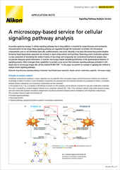
Nikon BioImaging Labs provide contract research services for microscope-based imaging and analysis to the biotech, pharma, and larger research communities. Each lab's full-service capabilities include access to cutting-edge microscopy instrumentation and software, but also the services of expert biologists and microscopists, who are available to provide quality cell culture, sample preparation, data acquisition, and data analysis services.
Search and filter over 125,000 Open Access Articles that utilize Nikon products and supported third party systems.
下载尼康显微镜产品相关的软件及固件程序。
找到最适于您工作流程的尼康物镜。
调整和操作精选尼康显微镜的步骤指南。
关于光学显微镜技术及其应用的网络研讨会。
通过尼康MicroscopyU网站为您介绍有关光学显微镜的教学资讯。
2023年10月
2022年6月
2022年2月
2021年12月
2021年6月
2021年4月
2021年1月