Nikon Europe B.V. | Europe & Africa
- pt Change Region
- Global Site
janeiro 2022
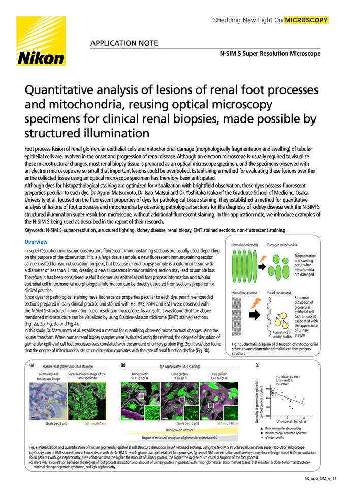
Foot process fusion of renal glomerular epithelial cells and mitochondrial damage (morphologically fragmentation and swelling) of tubular epithelial cells are involved in the onset and progression of renal disease. Although an electron microscope is usually required to visualize these microstructural changes, most renal biopsy tissue is prepared as an optical microscope specimen, and the specimens observed with an electron microscope are so small that important lesions could be overlooked. Establishing a method for evaluating these lesions over the entire collected tissue using an optical microscope specimen has therefore been anticipated.
Although dyes for histopathological staining are optimized for visualization with brightfield observation, these dyes possess fluorescent properties peculiar to each dye. Dr. Ayumi Matsumoto, Dr. Isao Matsui and Dr. Yoshitaka Isaka of the Graduate School of Medicine, Osaka University et al. focused on the fluorescent properties of dyes for pathological tissue staining. They established a method for quantitative analysis of lesions of foot processes and mitochondria by observing pathological sections for the diagnosis of kidney disease with the N-SIM S structured illumination super-resolution microscope, without additional fluorescent staining. In this application note, we introduce examples of the N-SIM S being used as described in the report of their research.