Nikon Europe B.V. | Europe & Africa
- pt Change Region
- Global Site
junho 2026
Confocal microscopy enables the visualization of the three-dimensional structures of biological samples with high spatial resolution, making it an indispensable tool in life science research. However, when observing thick biological samples, spherical aberration occurs due to the sample's thickness and the refractive index mismatch between the sample and the medium. Therefore, proper knowledge and techniques are required to eliminate distortion along the optical axis and accurately image the true 3D structure. In this application note, we demonstrate a method for achieving high-resolution 3D time-lapse imaging with minimized distortion deep within thick organoids.
Some objectives are equipped with a correction collar to compensate for variations in coverslip thickness as well as spherical aberration caused by sample thickness. This produces sharper images, particularly in the deeper sections of thicker samples, by minimizing spherical aberration. Standard usage involves focusing on the target in sample while rotating the correction collar to a position where the target appears brightest and at the highest resolution.
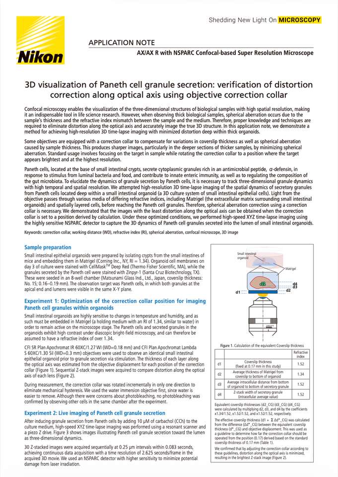
Paneth cells, located at the base of small intestinal crypts, secrete cytoplasmic granules rich in an antimicrobial peptide, α-defensin, in response to stimulus from luminal bacteria and food, and contribute to innate enteric immunity, as well as to regulating the composition of the gut microbiota. To elucidate the dynamics of granule secretion by Paneth cells, it is necessary to track three-dimensional granule dynamics with high temporal and spatial resolution. We attempted high-resolution 3D time-lapse imaging of the spatial dynamics of secretory granules from Paneth cells located deep within a small intestinal organoid (a 3D culture system of small intestinal epithelial cells). Light from the objective passes through various media of differing refractive indices, including Matrigel (the extracellular matrix surrounding small intestinal organoids) and spatially layered cells, before reaching the Paneth cell granules. Therefore, spherical aberration correction using a correction collar is necessary. We demonstrated that the images with the least distortion along the optical axis can be obtained when the correction collar is set to a position derived by calculation. Under these optimized conditions, we performed high-speed XYZ time-lapse imaging using the highly sensitive NSPARC detector to capture the 3D dynamics of Paneth cell granules secreted into the lumen of small intestinal organoids.
Keywords: correction collar, working distance (WD), refractive index (RI), spherical aberration, confocal microscope, 3D image